File list
From MicrobeWiki, the student-edited microbiology resource
This special page shows all uploaded files.
| Date | Name | Thumbnail | Size | User | Description | Versions |
|---|---|---|---|---|---|---|
| 22:22, 14 April 2024 | Screenshot 2024-04-14 at 6.22.01 PM.png (file) |  |
199 KB | Mccune2 | 1 | |
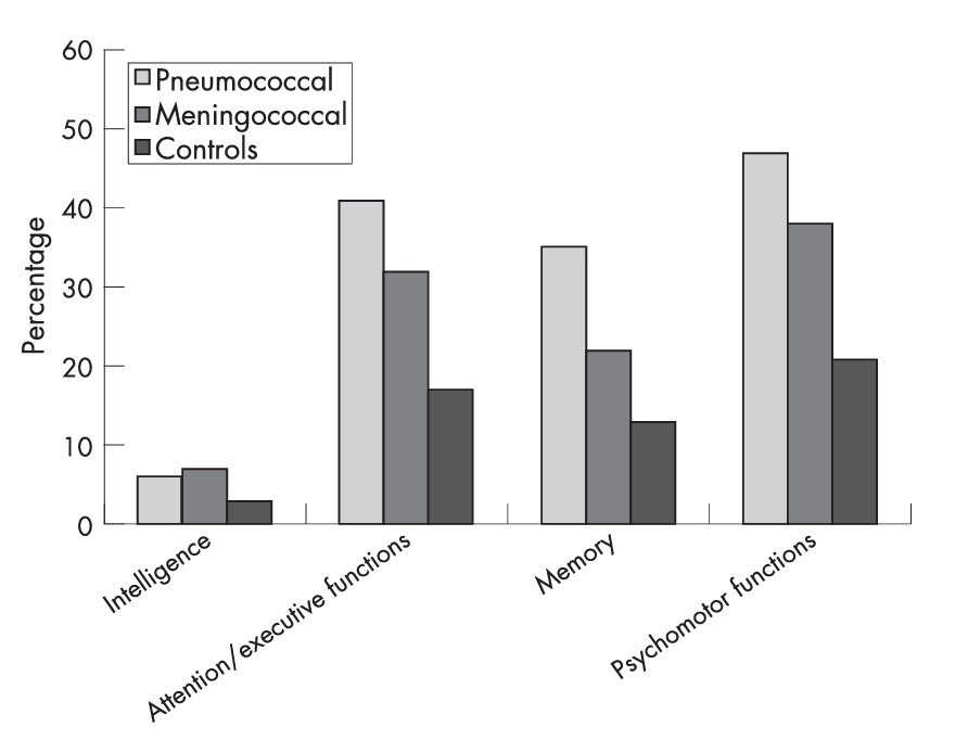
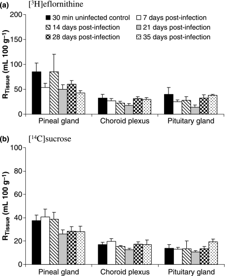
| 22:21, 14 April 2024 | Comparison graph.png (file) |  |
25 KB | Allen5 | 1 | |
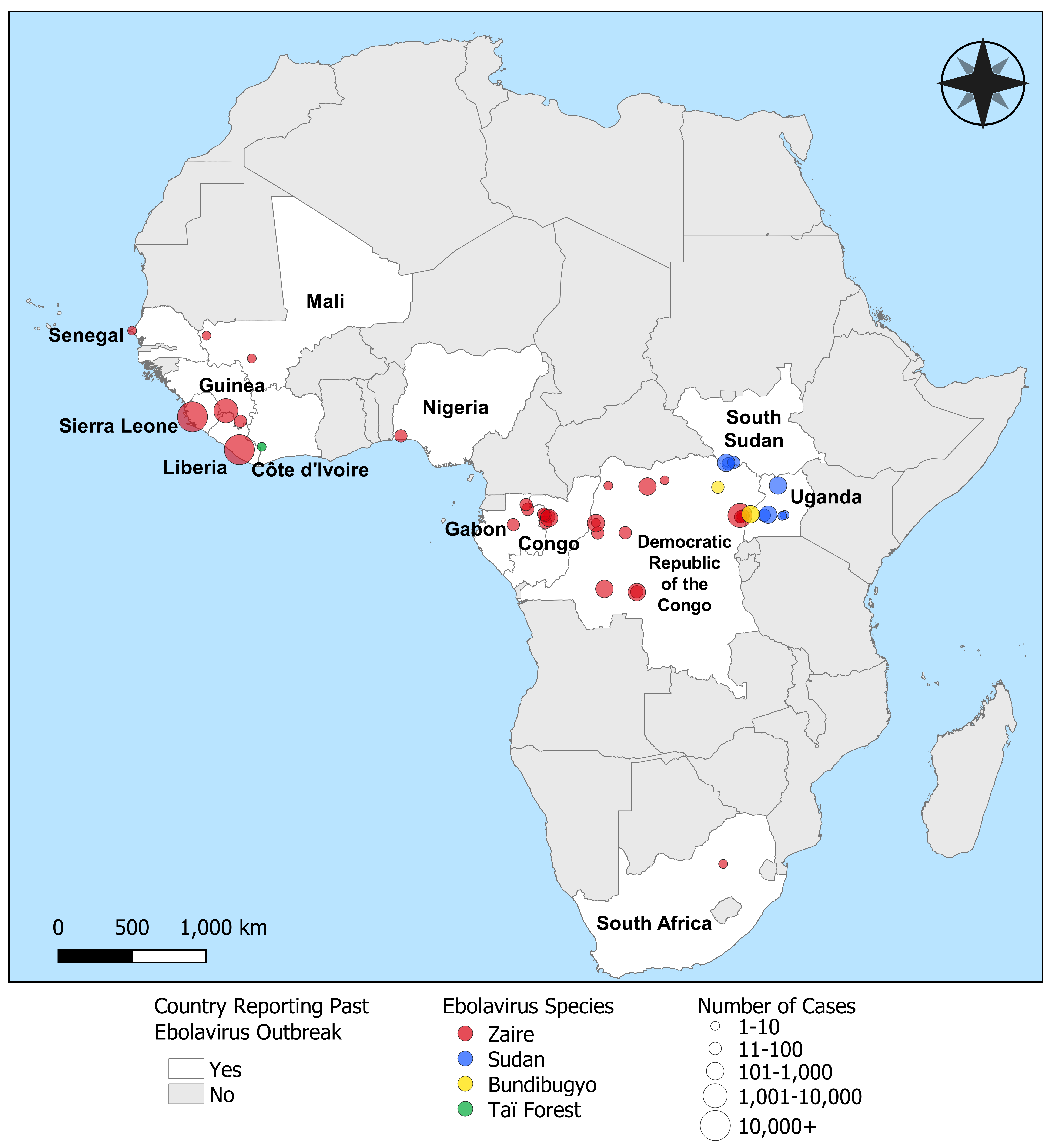
| 22:17, 14 April 2024 | Africanmap.png (file) | Error creating thumbnail: File with dimensions greater than 12.5 MP |
1.32 MB | Breard1 | 1 | |
| 22:13, 14 April 2024 | Screenshot 2024-04-14 at 6.09.29 PM.png (file) |  |
112 KB | Mccune2 | 1 | |
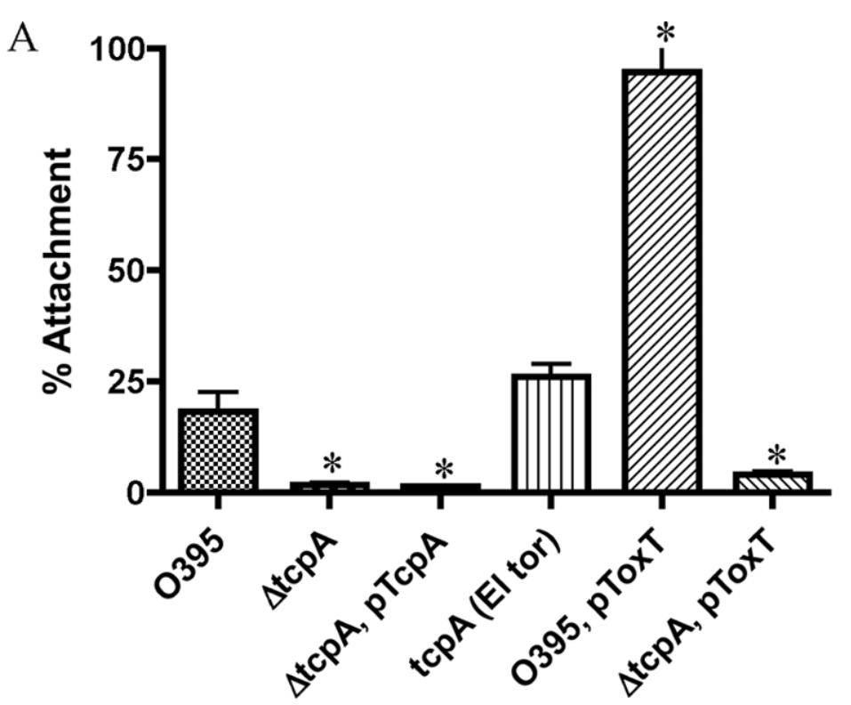
| 21:49, 14 April 2024 | TCP Attachment Experiment.png (file) |  |
156 KB | Raphael1 | 1 | |
| 21:46, 14 April 2024 | Screenshot 2024-04-14 at 5.38.29 PM.jpg (file) |  |
61 KB | Gest1 | 1 | |
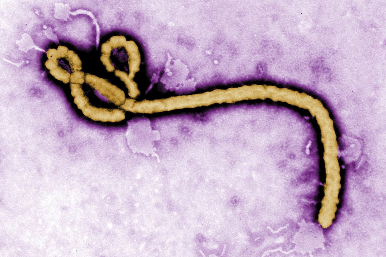
| 21:20, 14 April 2024 | Ebolabat.png (file) |  |
1.23 MB | Breard1 | 1 | |
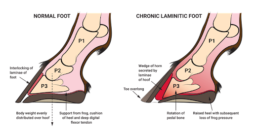
| 21:13, 14 April 2024 | Laminitis photo.png (file) |  |
87 KB | Allen5 | A diagram of a healthy vs. a laminitic horse hoof. | 1 |
| 21:09, 14 April 2024 | HTV genome.png (file) |  |
161 KB | Melo1 | 1 | |
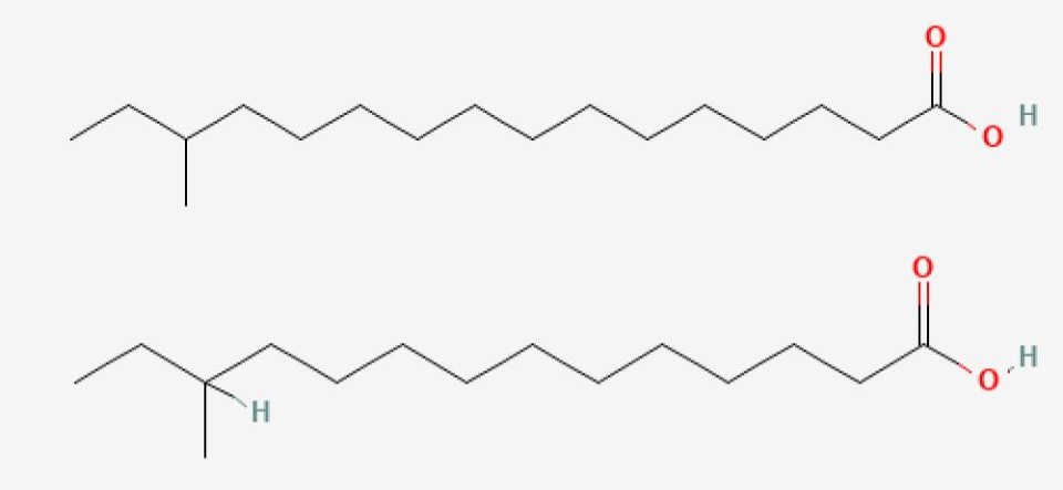
| 21:08, 14 April 2024 | Fatty acids.jpg (file) |  |
26 KB | Pardue | 1 | |
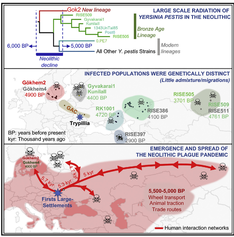
| 21:01, 14 April 2024 | Graphical Abstract Rascovan et al .png (file) |  |
661 KB | Schwingel | Graphical abstract describing the hypothesis of a Neolithic Yersinia pestis plague. Image from Rascovan, Nicolás, Karl-Göran Sjögren, Kristian Kristiansen, Rasmus Nielsen, Eske Willerslev, Christelle Desnues, and Simon Rasmussen. “Emergence and Spread of Basal Lineages of Yersinia Pestis during the Neolithic Decline.” 2019. Cell 176 (1–2): 295-305.e10. | 1 |
| 20:45, 14 April 2024 | Crohns dreamstime s 119957874.jpg (file) |  |
228 KB | Arone1 | 1 | |
| 20:44, 14 April 2024 | Palcam listeria.jpg (file) |  |
203 KB | Pardue | 1 | |
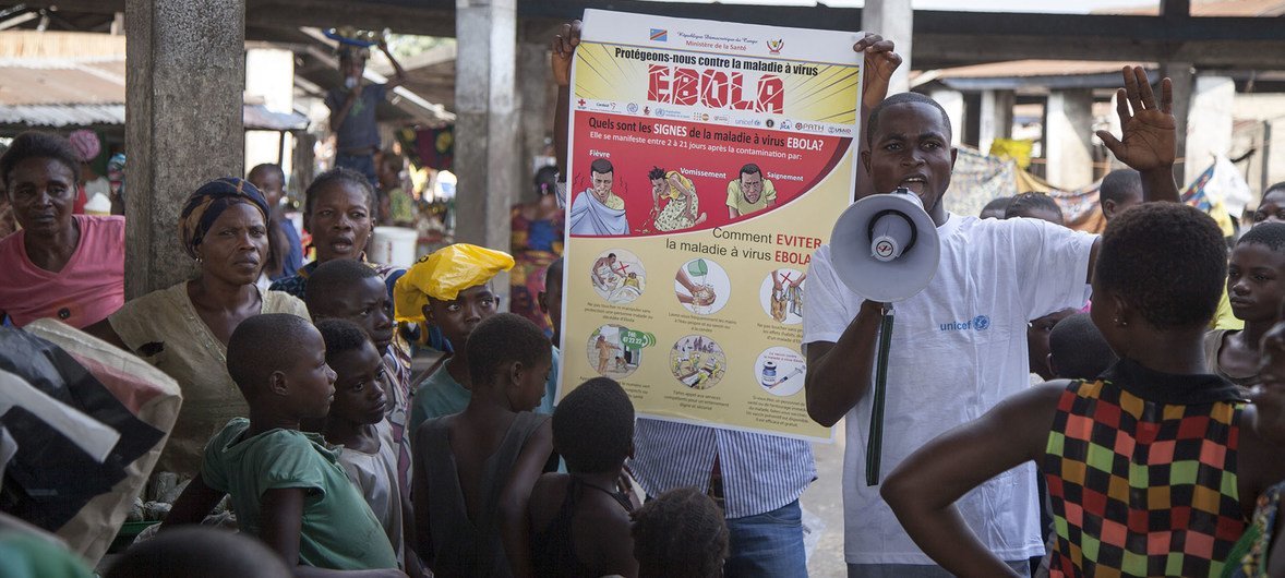
| 20:37, 14 April 2024 | UNICEF.jpg (file) |  |
146 KB | Breard1 | 1 | |
| 20:30, 14 April 2024 | Screen Shot 2024-04-14 at 4.29.26 PM.png (file) |  |
447 KB | Breard1 | 1 | |
| 20:25, 14 April 2024 | Matt1.jpg (file) |  |
156 KB | Nguyen | 1 | |
| 20:15, 14 April 2024 | EBOVtable.png (file) |  |
73 KB | Breard1 | 1 | |
| 19:57, 14 April 2024 | Topical.webp (file) |  |
7 KB | Lydon1 | 1 | |
| 19:56, 14 April 2024 | Circular representation of genomes of M. haemocanis and M. haemofelis.webp (file) |  |
99 KB | Erbyers | 1 | |
| 19:47, 14 April 2024 | Microbiome Image.png (file) |  |
187 KB | Lydon1 | 1 | |
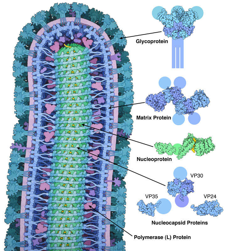
| 19:46, 14 April 2024 | Ebolablue.jpg (file) |  |
232 KB | Breard1 | 1 | |
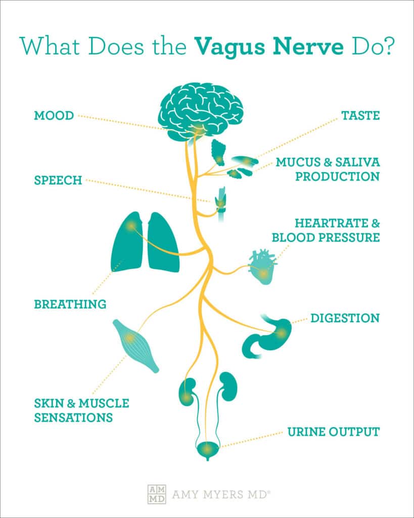
| 19:38, 14 April 2024 | Vagus Nerve Innervation.jpg (file) |  |
42 KB | Lee10 | 1 | |
| 19:34, 14 April 2024 | Lactobacillus Treatment.png (file) |  |
151 KB | Lee10 | 1 | |
| 19:26, 14 April 2024 | Vagus Nerve and Gut Microbiome.jpg (file) |  |
8 KB | Lee10 | 1 | |
| 19:23, 14 April 2024 | CBT.jpg (file) |  |
31 KB | Lee10 | 1 | |
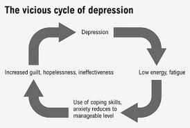
| 19:19, 14 April 2024 | Depression Cycle.jpg (file) |  |
7 KB | Lee10 | 1 | |
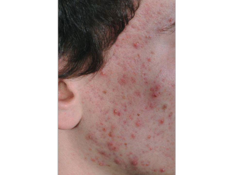
| 19:18, 14 April 2024 | Acne Image.jpg (file) |  |
41 KB | Lydon1 | 1 | |
| 19:15, 14 April 2024 | Neurotransmitter Release.jpg (file) | 100 KB | Lee10 | 1 | ||
| 19:09, 14 April 2024 | Ribavirin.jpeg (file) |  |
69 KB | Melo1 | 1 | |
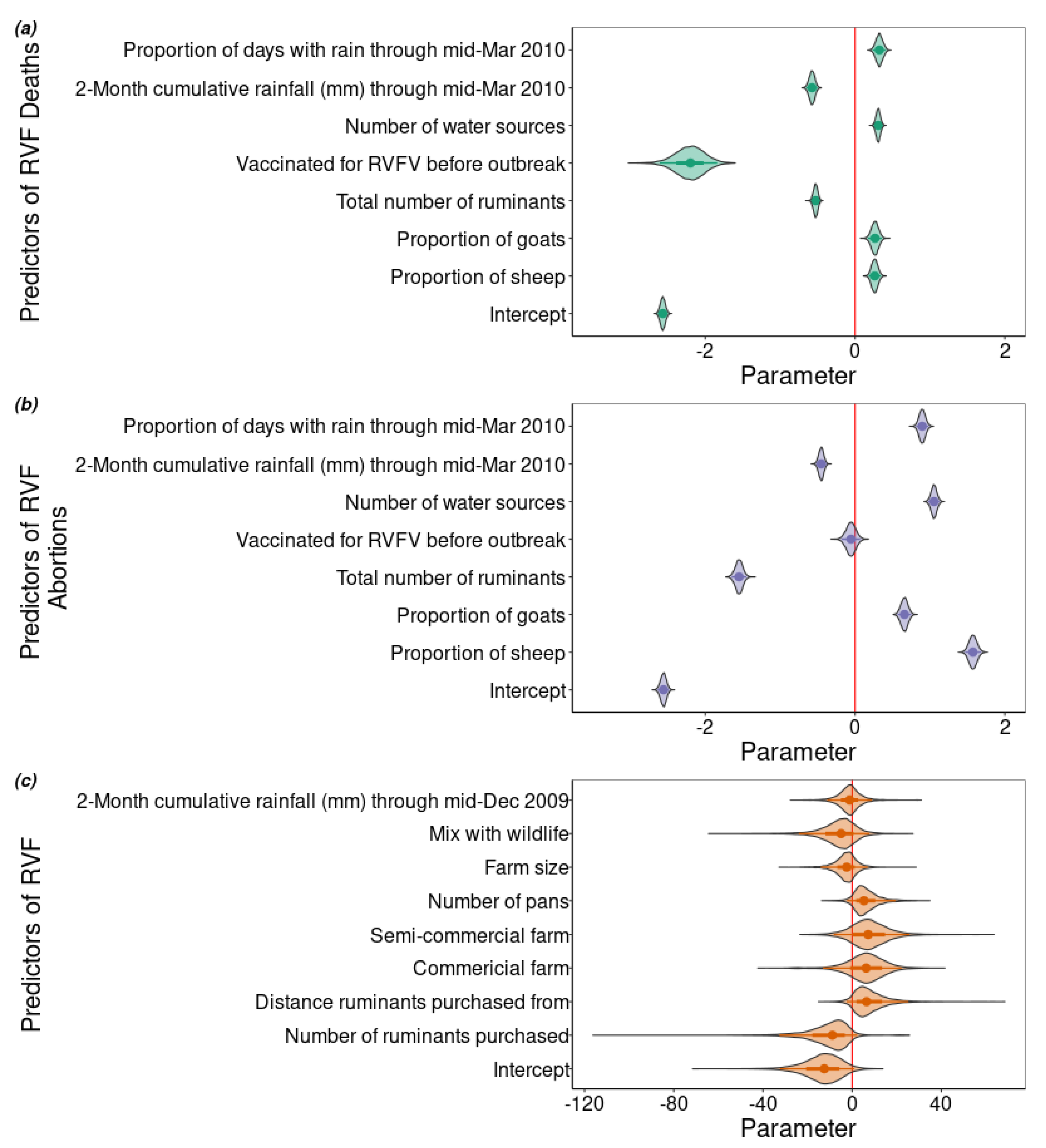
| 19:05, 14 April 2024 | Predictors of RVF.png (file) |  |
488 KB | Caine1 | 1 | |
| 19:03, 14 April 2024 | Sin Nombre University of New Mexico Hospital 1993.jpg (file) |  |
77 KB | Melo1 | 1 | |
| 18:57, 14 April 2024 | Liu Chart.jpeg (file) |  |
76 KB | Lydon1 | 1 | |
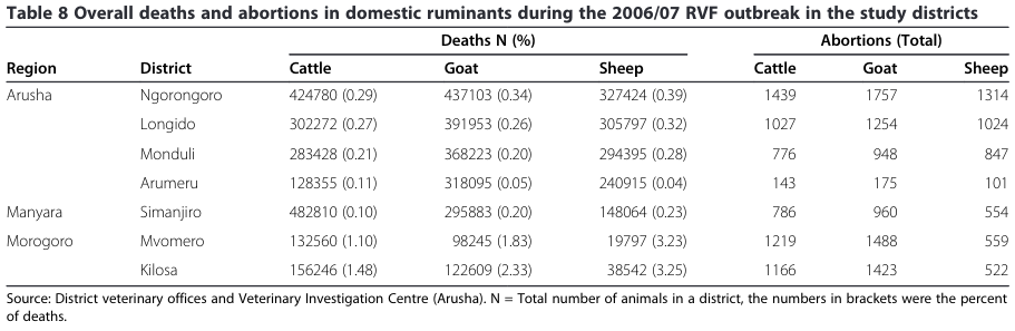
| 18:38, 14 April 2024 | RVF-2007-death-abortions.png (file) |  |
72 KB | Caine1 | 1 | |
| 18:36, 14 April 2024 | Ebolapurpleimage.jpg (file) |  |
204 KB | Breard1 | 1 | |
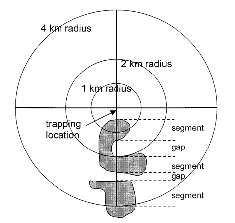
| 18:24, 14 April 2024 | 2001 Landscape structure influences continental distribution of hantavirus in deer mice, Figure 1.png (file) |  |
61 KB | Melo1 | 1 | |
| 17:59, 14 April 2024 | RVFV.jpg (file) |  |
82 KB | Caine1 | 1 | |
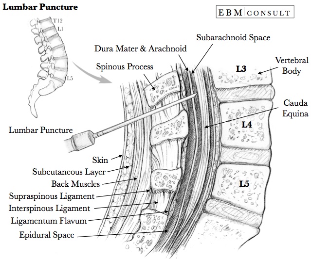
| 17:41, 14 April 2024 | -223 KB- Lumbar Puncture Procedure Image - EBM Consult.jpg (file) |  |
135 KB | Gest1 | 1 | |
| 17:13, 14 April 2024 | Mycoplasma haemocanis infection, canine blood smear - Merck Veterinary Manual.jpg (file) |  |
102 KB | Erbyers | 1 | |
| 16:05, 14 April 2024 | HAT mice.jpg (file) |  |
76 KB | Mclaughlin2 | 1 | |
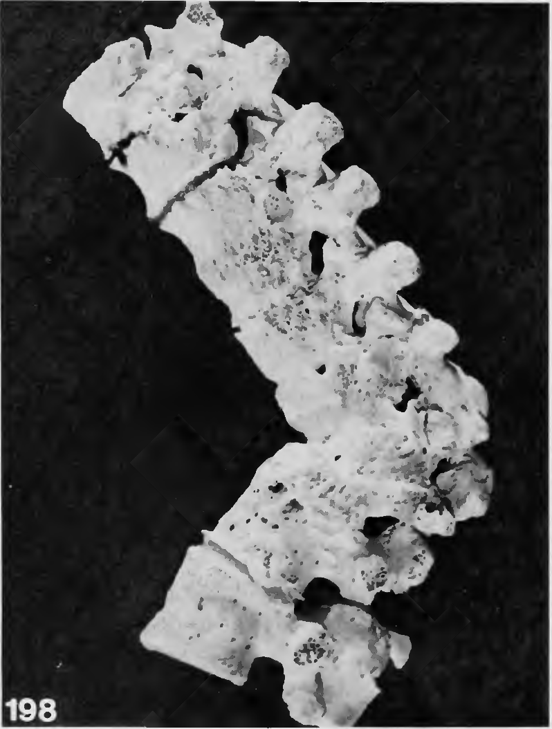
| 15:44, 14 April 2024 | Tuberculosis of Spine Buikstra.png (file) |  |
595 KB | Schwingel | Lateral view of spine affected by tuberculosis, partly healed, affecting thoracic vertebrae 7 and 8 and first lumbar vertebra. Image from Buikstra, J.E. ed. "Ortner's identification of pathological conditions in human skeletal remains." 2019. | 1 |
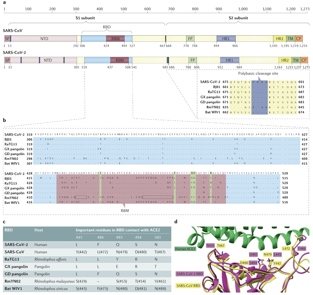
| 15:41, 14 April 2024 | COVID.png (file) |  |
1.21 MB | Yang | 1 | |
| 15:37, 14 April 2024 | HAT clothing.jpg (file) |  |
554 KB | Mclaughlin2 | 1 | |
| 15:31, 14 April 2024 | HAT treatment.gif (file) |  |
32 KB | Mclaughlin2 | 1 | |
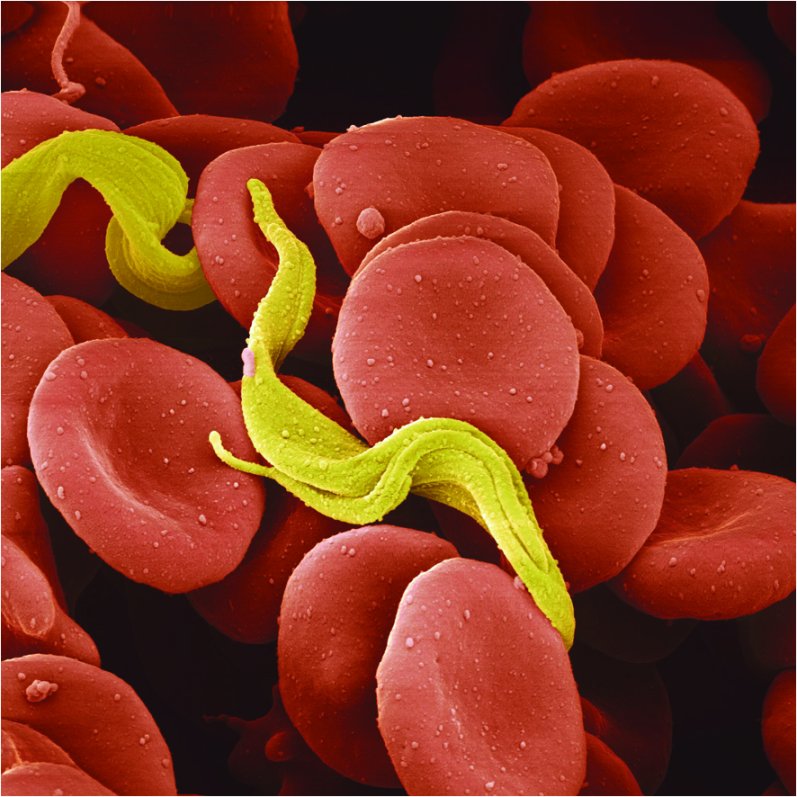
| 15:08, 14 April 2024 | T.Brucei.jpg (file) |  |
112 KB | Mclaughlin2 | 1 | |
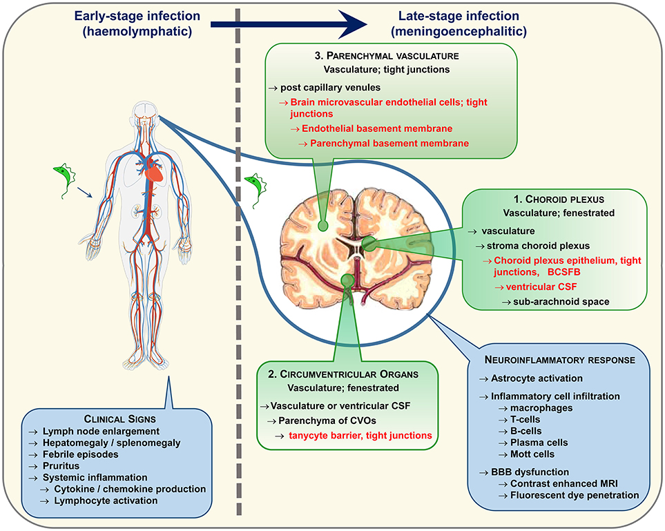
| 15:00, 14 April 2024 | HAT path.jpg (file) |  |
369 KB | Mclaughlin2 | 1 | |
| 14:50, 14 April 2024 | Humanastro.jpg (file) |  |
120 KB | Ehaggin | 1 | |
| 14:50, 14 April 2024 | HAT symptoms.jpg (file) |  |
534 KB | Mclaughlin2 | 1 | |
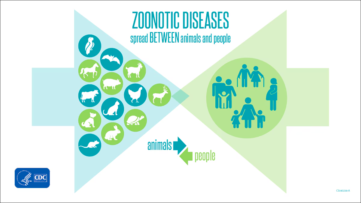
| 14:47, 14 April 2024 | Zoonoses.png (file) |  |
343 KB | Caine1 | 1 | |
| 14:41, 14 April 2024 | Phytotherapy.jpg (file) |  |
308 KB | Newman5 | 1 | |
| 14:30, 14 April 2024 | HAT distribution.jpg (file) |  |
1.7 MB | Mclaughlin2 | 1 |
