Unused files
From MicrobeWiki, the student-edited microbiology resource
The following files exist but are not embedded in any page. Please note that other web sites may link to a file with a direct URL, and so may still be listed here despite being in active use.
Showing below up to 250 results in range #251 to #500.
 US-Diphtheria.gif 770 × 512; 8 KB
 M.j..gif 225 × 225; 58 KB
 Water supply.jpg 700 × 460; 143 KB
 Methanococcus.jpg 300 × 200; 31 KB
 A02 bacteria full.jpg 518 × 656; 374 KB
 P01.jpg 240 × 162; 6 KB
 Hyphomonas.jpg 303 × 228; 11 KB
 Hyphomonasgenome.jpg 135 × 135; 4 KB
 Halorhodospira.gif 193 × 440; 71 KB
 Yeast.jpg 500 × 500; 160 KB
 240px-Streptococcus mutans 01.jpg 240 × 175; 12 KB
 240px-streptococcus mutans 01.jpg 240 × 175; 12 KB
 Picture 1.jpg 240 × 210; 35 KB
 OTSM.jpg 447 × 500; 77 KB
 Pg03c-1-.jpg 153 × 145; 10 KB
 Staphylococcus epidermidis lores.jpg 700 × 412; 55 KB
 Sporothrix 1.jpg 596 × 401; 46 KB
 Figure 1.JPG 935 × 1,209; 70 KB
 Periodontitis-1-.gif 226 × 227; 27 KB
 HCgramstain.jpg 150 × 100; 4 KB
 HCgramstain01.jpg 1,225 × 816; 82 KB
 HC02.JPG 640 × 400; 21 KB
 HC20C.JPG 640 × 400; 15 KB
 HC37C.JPG 640 × 400; 21 KB
 S.saprophyticus genome.png 600 × 600; 100 KB
 DennisKunkelMicroscopy,Inc.jpg 275 × 367; 61 KB
 C immitis structure.png 300 × 204; 123 KB
 26644C.jpg 275 × 324; 121 KB
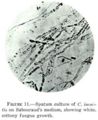 Coccidioides immitis on Sabouraud's medium.jpg 301 × 369; 45 KB
 Lactobacillus delbrueckii.jpg 72 × 48; 16 KB
 L delbrueckii subsp bulgaricus.jpg 72 × 48; 15 KB
 Stanford.jpg 311 × 149; 7 KB
 Meso 2.jpg 100 × 125; 10 KB
 Image-C albicans en.jpg 640 × 480; 89 KB
 A.b.jpg 133 × 127; 3 KB
 Hmk.jpg 133 × 127; 3 KB
 96546F.jpg 349 × 275; 92 KB
 Chromosomes.jpg 480 × 320; 62 KB
 Aa microscope8.jpg 210 × 208; 10 KB
 DroppedImage 3.png 187 × 161; 28 KB
 Pdidemni cells.png 227 × 161; 50 KB
 Lpatella.png 241 × 161; 86 KB
 Alfalfa.JPG 300 × 202; 10 KB
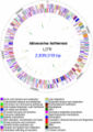 GenomeIloihiensis.jpg 400 × 566; 64 KB
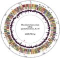 Map K10 Genome.jpg 440 × 421; 48 KB
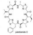 Patellamide.jpg 206 × 217; 7 KB
 Miniproch.jpg 62 × 68; 2 KB
 Amebiasis LifeCycle.gif 435 × 548; 28 KB
 Thermus Thermophilus.jpg 200 × 168; 23 KB
 Thermus.jpg 200 × 133; 59 KB
 Tsetse haemocyte and S glossinidius.jpg 473 × 290; 15 KB
 Tsetse haemocyte and S glossinidius (2).jpg 375 × 266; 13 KB
 Rickettsia rickettsii3..gif 252 × 198; 11 KB
 Rickettsia rickettsii3.jpg.gif 252 × 198; 11 KB
 Rickettsia rickettsii3.jpg 252 × 198; 24 KB
 C immitis endospores.jpg 700 × 472; 30 KB
 Rhizobium etli bacteria.JPG 216 × 158; 11 KB
 Rhizobium etli nitrogen fixing nodules.JPG 282 × 163; 11 KB
 Pyris.jpg 155 × 246; 30 KB
 Zh40090433050006.jpeg 774 × 593; 190 KB
 Desulfovibrio.jpg 170 × 170; 8 KB
 Streptomycesgriseus.jpg 142 × 201; 13 KB
 Toothdecay.png 312 × 651; 242 KB
 S avermitilis.jpg 274 × 406; 79 KB
 S avermitilis genome.png 600 × 600; 186 KB
 River blindness.jpg 210 × 300; 10 KB
 Thermotoga neapolitana edited.JPG 2,083 × 1,250; 78 KB
 Wiki-1.jpg 630 × 194; 50 KB
 Leprae.jpg 411 × 258; 20 KB
 Zoogloea.jpg 403 × 260; 21 KB
 Zoogloea ramigera.jpg 403 × 260; 21 KB
 Micro pic.jpg 128 × 90; 4 KB
 Wiki5.jpg 369 × 464; 29 KB
 Ex 1.jpg 250 × 163; 8 KB
 Octopus spring.jpg 960 × 720; 54 KB
 Octopus spring.png 962 × 600; 778 KB
 BChlc.png 436 × 342; 55 KB
 Photo.png 677 × 612; 478 KB
 Whiterot.gif 640 × 480; 111 KB
 OilSheenFromValdezSpill.jpg 384 × 256; 15 KB
 PAH.jpg 448 × 332; 27 KB
 Ncycl.jpg 373 × 232; 82 KB
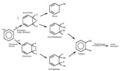 PAH degradation.jpg 448 × 266; 13 KB
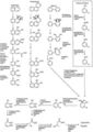 PCB degradation.jpg 317 × 448; 28 KB
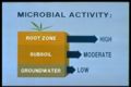 Microbial activity.jpg 384 × 256; 16 KB
 Simplencycle.gif 515 × 388; 6 KB
 Greehouse Effect.jpg 490 × 391; 214 KB
 CCycle.gif 450 × 486; 61 KB
 DCP 7175.JPG 396 × 266; 22 KB
 500px-Troph flowchart.svg.png 500 × 390; 27 KB
 Anaerobic decomposition.PNG 641 × 469; 165 KB
 Methane pic.jpg 450 × 600; 73 KB
 Image-File.jpg 450 × 600; 73 KB
 Humus.jpg 130 × 97; 3 KB
 Gley.jpg 640 × 426; 118 KB
 Presentation1.jpg 960 × 720; 37 KB
 Enterobacteriaceae.jpg 587 × 319; 28 KB
 Presentation2.jpg 960 × 720; 23 KB
 Bacteria2.jpg 500 × 400; 21 KB
 Hemicellulose.jpg 960 × 720; 33 KB
 HemicelluloseDecomp.jpg 960 × 720; 33 KB
 Acidobacteria.jpg 250 × 200; 27 KB
 Art3-t.jpg 350 × 495; 107 KB
 Oparin.jpg 415 × 300; 29 KB
 Cartoon.jpg 124 × 109; 4 KB
 Amc0839l.jpg 400 × 351; 22 KB
 Rhizobact.jpg 632 × 422; 364 KB
 Phytophtora reproduction.png 685 × 600; 315 KB
 Life Cycle.png 409 × 262; 51 KB
 Phytophora.jpg 321 × 277; 60 KB
 Ahsan P1.png 252 × 199; 102 KB
 Picture2.jpg 1,115 × 368; 63 KB
 Phytophthora life cycle.png 409 × 262; 51 KB
 Potato1.jpg 380 × 285; 16 KB
 Potato2.jpg 380 × 285; 19 KB
 Spores.gif 454 × 746; 212 KB
 Tomato.jpg 148 × 150; 10 KB
 Zoospore.jpg 524 × 372; 73 KB
 Vcholera.jpg 600 × 861; 98 KB
 Acid2.jpg 200 × 133; 29 KB
 VAGINA.gif 430 × 399; 21 KB
 NEA04.gif 450 × 407; 92 KB
 Saltwater aquarium.jpg 500 × 331; 49 KB
 Miso.gif 450 × 304; 101 KB
 Brackish.jpg 736 × 450; 108 KB
 Stinkytofu.jpg 400 × 598; 33 KB
 Ear picture.gif 650 × 515; 86 KB
 Showercurtain.jpg 354 × 354; 17 KB
 Cleaningmoney.jpg 298 × 181; 12 KB
 Lakemagadi.png 360 × 270; 148 KB
 Rumen 3.jpg 530 × 770; 99 KB
 Source pH.jpg 636 × 386; 12 KB
 2001111.jpg 500 × 333; 29 KB
 PH.jpg 636 × 386; 12 KB
 Microbetable1.jpg 671 × 359; 70 KB
 Myco.gif 200 × 150; 30 KB
 Bugs.jpg 320 × 237; 56 KB
 RunOff.jpg 500 × 375; 169 KB
 Underwater McMurdo Sound Ice Wall.JPG 450 × 337; 37 KB
 Aurora Australis Antarctica Amundsen-Scott South Pole Station.JPG 640 × 427; 28 KB
 Villi em.jpg 228 × 141; 15 KB
 Microvilli.jpg 216 × 175; 17 KB
 CALKINGLACIER.JPG 440 × 330; 63 KB
 Mud Volcano.jpg 500 × 800; 138 KB
 EchinusGeyser.jpg 160 × 120; 23 KB
 Fresh Aq.jpg 700 × 525; 73 KB
 Vostok main.jpg 450 × 360; 19 KB
 Aquarium.jpg 600 × 450; 98 KB
 Scenic 1000 coronation18.jpg 475 × 315; 78 KB
 DONJUAN.JPG 450 × 338; 33 KB
 SPINERIDGE.JPG 450 × 338; 24 KB
 GERLACHESTRAIT5.JPG 450 × 336; 43 KB
 PALMERICECAVE.JPG 450 × 338; 35 KB
 SUNANDDISCOVERY.JPG 450 × 302; 16 KB
 GLACIERANDMOUNTAINS.JPG 450 × 295; 40 KB
 ICEFALL2.JPG 450 × 299; 31 KB
 NACREOUSCLOUDS3.JPG 450 × 352; 8 KB
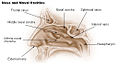 Illu nose nasal cavities.jpg 520 × 300; 39 KB
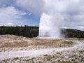 Old Faithful Geyser.jpg 246 × 184; 17 KB
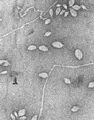 Virus in Yellowstone.jpg 154 × 197; 12 KB
 Yellowstone Hot Spring.jpg 263 × 165; 16 KB
 Donlan1b.jpg 500 × 339; 43 KB
 K9481-1.jpg 411 × 640; 121 KB
 Cinder.JPG 650 × 431; 46 KB
 Porteous1.jpg 372 × 420; 121 KB
 Porteous2.jpg 373 × 432; 124 KB
 Porteous3.jpg 367 × 401; 144 KB
 Large Intestine.jpg 216 × 158; 10 KB
 Chicken.jpg 360 × 242; 60 KB
 City Champs 1.jpg 604 × 453; 69 KB
 Gram-Stained Bifido-1543654.jpg 216 × 158; 10 KB
 BacterialIsolates.png 255 × 327; 27 KB
 Dock.jpg 1,024 × 768; 309 KB
 Hayyyy.jpg 273 × 179; 26 KB
 Yak.jpg 392 × 252; 27 KB
 PHIL 1187 lores.jpg 699 × 472; 49 KB
 Bacteriumonprotist1.JPG 347 × 220; 10 KB
 Bacteriumoncellulose1.JPG 341 × 255; 16 KB
 10043 lores.jpg 700 × 491; 63 KB
 964 lores.jpg 700 × 468; 95 KB
 COW.jpg 640 × 449; 102 KB
 Methanobacterium formicicum.jpg 1,376 × 913; 77 KB
 Fistulatedcows2cf8.jpg 580 × 435; 33 KB
 Fistulated cow.jpg 443 × 292; 15 KB
 Pyarmid.JPG 1,104 × 448; 60 KB
 Mobile.jpg 2,687 × 1,800; 217 KB
 Staphyylococci.jpg 700 × 467; 46 KB
 Mail-2.jpg 347 × 220; 35 KB
 Mail-212.jpg 347 × 220; 35 KB
 D383-2i.jpg 252 × 165; 26 KB
 Grass,jpg.jpg 400 × 400; 125 KB
 Newwater.jpg 330 × 177; 11 KB
 Closefist2.jpg 443 × 292; 15 KB
 Teeth.JPG 450 × 470; 30 KB
 SceneryNorrisGeyserBasin.jpg 360 × 240; 24 KB
 Kenya.jpg 563 × 458; 58 KB
 Throat.JPG 400 × 320; 22 KB
 Gums.JPG 490 × 308; 27 KB
 Actinomycetes6-small.jpeg 230 × 338; 25 KB
 16902.jpg 1,999 × 1,277; 661 KB
 AN.JPG 291 × 202; 6 KB
 Pseudomonas aeruginosa - Image.jpg 124 × 97; 4 KB
 Biofilm2.jpg 313 × 208; 6 KB
 Run Off.jpg 211 × 157; 18 KB
 Thermoplasma acidophilum.jpg 100 × 112; 11 KB
 Tongue.jpg 1,196 × 625; 166 KB
 5 days.jpg 395 × 336; 28 KB
 Haemophilus influenzae fig1.jpg 582 × 398; 54 KB
 Burkholderia cepacia.jpg 700 × 475; 69 KB
 Peyer.jpg 256 × 99; 4 KB
 Danny.jpg 527 × 342; 36 KB
 TaqPol.jpg 180 × 180; 8 KB
 Moraxella catarrhalis.jpg 470 × 282; 21 KB
 Helicobacter pylori.jpg 175 × 260; 13 KB
 Stomachulcers.jpg 184 × 230; 51 KB
 Sargassumweeds.jpg 600 × 450; 110 KB
 Synechococcus TEM.jpg 120 × 116; 8 KB
 6851 01.jpg 240 × 301; 16 KB
 H. pylori.png 216 × 195; 35 KB
 PentosePhosphatePathway-OxidativeNADPH.PNG 983 × 106; 8 KB
 Ulcer!.jpg 400 × 408; 28 KB
 Cladosporium.jpg 275 × 343; 101 KB
 Cryptosporidium.jpg 349 × 275; 67 KB
 Giardia.jpg 275 × 343; 73 KB
 Penicillium.jpg 349 × 275; 115 KB
 Scopulariposis.jpg 275 × 324; 49 KB
 Sneeze.jpg 520 × 359; 38 KB
 Lichen.jpg 384 × 257; 35 KB
 Corynebacterium ulcerans 01.jpg 700 × 476; 32 KB
 300px-Isolated bacteria - Micrococcus luteus.jpg 800 × 600; 73 KB
 Horse2.jpg 300 × 308; 24 KB
 Untitled1.png 241 × 56; 10 KB
 Witwatersrand basin.jpg 134 × 101; 3 KB
 Honeybee.jpg 194 × 252; 88 KB
 Rpv1.JPG 648 × 472; 76 KB
 600px-Serratia marcescens 01.jpg 600 × 599; 44 KB
 HTLV genome.jpg 595 × 257; 30 KB
 Pic55525.jpg 640 × 480; 58 KB
 Image for wiki.jpg 300 × 300; 12 KB
 B bacilliformis genome.png 600 × 600; 97 KB
 Solo-bacteria.jpg 220 × 151; 15 KB
 SSV infection.jpg 375 × 362; 73 KB
 Bluebl1.jpg 225 × 212; 30 KB
 Slide1.JPG 960 × 720; 18 KB
 Gsb1 bacteria.jpg 537 × 720; 21 KB
 Fig. 2 Origins.JPG 660 × 512; 31 KB
 Chiralsynthesis.JPG 660 × 512; 31 KB
 CPL.JPG 762 × 512; 42 KB
 Slide2.JPG 537 × 720; 71 KB
 Slide3.JPG 537 × 720; 43 KB


.jpg)


