Unused files
From MicrobeWiki, the student-edited microbiology resource
The following files exist but are not embedded in any page. Please note that other web sites may link to a file with a direct URL, and so may still be listed here despite being in active use.
Showing below up to 250 results in range #51 to #300.
 T namibiensis1.jpg 550 × 477; 81 KB
 Vsd1 c.gif 500 × 395; 158 KB
 Vvpath.jpg 600 × 430; 31 KB
 Wiggles3.jpg 363 × 510; 74 KB
 Vsdcrename.jpg 500 × 395; 67 KB
 Beard6b.gif 620 × 360; 10 KB
 Diseases.gif 864 × 576; 351 KB
 FDRFalls.jpg 600 × 401; 22 KB
 X.medited.gif 321 × 238; 68 KB
 Diseasedplant.gif 461 × 420; 132 KB
 Tomatovictim.gif 355 × 241; 62 KB
 Strep mycelium.gif 720 × 488; 283 KB
 Colony.gif 302 × 297; 80 KB
 Biofilm.JPG 301 × 332; 43 KB
 Archaeoglobus 1.gif 283 × 161; 39 KB
 Biofilm.gif 301 × 332; 109 KB
 Archaeoglobus 2.gif 232 × 256; 62 KB
 Skull.jpg 435 × 648; 45 KB
 Methanoculleus 1.JPG 352 × 310; 52 KB
 Methanopyrus 1.jpg 285 × 267; 17 KB
 Methanopyrus 2.jpg 432 × 132; 8 KB
 Methanothermobacter 1.gif 231 × 198; 50 KB
 Magadi.gif 354 × 255; 81 KB
 Magadi 2.jpg 119 × 180; 14 KB
 Viralbiorealm banner2.jpg 800 × 150; 60 KB
 Thermoproteus.jpg 498 × 315; 29 KB
 Em ttv1.gif 640 × 476; 48 KB
 P8100450 mammoth spring large.jpg 600 × 800; 141 KB
 23295A.jpg 275 × 344; 59 KB
 395-10.jpg 718 × 600; 110 KB
 Headbg.jpg 1,941 × 220; 53 KB
 Siphoviridae.jpg 649 × 613; 135 KB
 Tectiviridae.jpg 752 × 600; 62 KB
 Leviviridae.jpg 738 × 595; 55 KB
 Potyvirus.jpg 1,038 × 768; 236 KB
 Guttaviridae.jpg 476 × 480; 163 KB
 Huitlacoche.jpg 200 × 359; 18 KB
 Virus.png 400 × 350; 141 KB
- Error creating thumbnail: File missingRhodospirillum rubrum.jpg 300 × 194; 20 KB
 Whipplei1.jpg 300 × 300; 31 KB
 Whipplei2.jpg 500 × 500; 52 KB
 Thermopl.gif 567 × 634; 366 KB
 BIFIDO2P.jpg 93 × 144; 4 KB
 BIFIDO2P1.jpg 144 × 93; 4 KB
 Ftularensis.jpg 500 × 394; 16 KB
 Ticktransmission.jpg 606 × 222; 18 KB
 Infectionspread.jpg 606 × 594; 55 KB
 Haemolyticus main pics.jpg 200 × 270; 18 KB
 Sullivan wolbachia.jpg 250 × 270; 23 KB
 Wolbachia genome.jpg 300 × 263; 88 KB
 She p.jpg 260 × 260; 60 KB
 Shewanella loihica.jpg 260 × 260; 60 KB
 060718 gold bacteria 01.jpg 277 × 221; 53 KB
 060802103513.jpg 300 × 229; 18 KB
 Bbsem.jpeg 320 × 235; 50 KB
 Metabolism.jpeg 967 × 1,280; 321 KB
 Bt1.jpeg 249 × 174; 21 KB
 Pic 1.jpg 275 × 343; 75 KB
 Chlamydophila abortus.gif 300 × 200; 22 KB
 Bt2.jpeg 269 × 186; 5 KB
 Bt3.JPG 1,011 × 113; 12 KB
 Corynebacterium glutamicum.png 354 × 261; 69 KB
 Bt4.JPG 201 × 255; 11 KB
 Chromohalobacter Salexigens BacMaps.png 600 × 600; 110 KB
 Pyrobaculum islandicum.JPG 515 × 338; 37 KB
 Pyrobaculum islandicum.jpg 515 × 338; 37 KB
 Bacteriaaaaaa.jpg 416 × 366; 25 KB
 Diversity.jpg 300 × 200; 35 KB
 Nrmicro1264-f3.jpg 600 × 614; 33 KB
 D.radiodurans.r1dna.jpg 284 × 256; 7 KB
 Bimm120 Vibrio vulnificus pic.jpg 580 × 435; 35 KB
 180px-Saccharum officinarum.jpg 180 × 240; 14 KB
 QFEVER 1.jpg 100 × 71; 21 KB
 Dehalococcoides ethenogenes.jpg 300 × 199; 28 KB
 DE2.jpg 600 × 313; 40 KB
 800px-Campylobacter jejuni 01.jpg 800 × 544; 12 KB
 Willi copy.jpg 700 × 509; 99 KB
 Gloeobacter violaceus.jpg 100 × 135; 9 KB
 JCS01382F1.jpg 512 × 413; 11 KB
 240px-Campylobacter jejuni 01.jpg 240 × 163; 2 KB
 JCS01382F8.gif 440 × 416; 19 KB
 Pic.jpg 124 × 123; 5 KB
 Pprofundum SS9.nfp.png 674 × 476; 199 KB
 Geobacterflag.jpg 250 × 239; 21 KB
 Micoplasma2.gif 300 × 200; 43 KB
 Pic2.gif 300 × 399; 83 KB
 Alactobacillus.jpg 163 × 157; 25 KB
 Rlv3841chrom.jpg 600 × 577; 155 KB
 Pelo1.JPG 214 × 179; 6 KB
 Pelo2.JPG 250 × 250; 16 KB
 Nitrification.gif 180 × 142; 5 KB
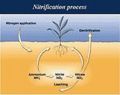 Nitrification.JPG 180 × 142; 7 KB
 Methylococcus capsulatus str. Bath.jpg 300 × 200; 35 KB
 B.mallei1.jpg 510 × 466; 53 KB
 V cholerae1.jpg 205 × 113; 6 KB
 Leifson flagella stain.jpg 419 × 341; 48 KB
 Gram stain 2.jpg 420 × 434; 27 KB
 Magnetotactic bacterium2 .jpg 240 × 234; 4 KB
 Metabilic Pathways.gif 1,332 × 1,022; 51 KB
 Matabolic Pathways.gif 1,332 × 1,022; 51 KB
 Image of thermocellum interior.jpg 300 × 200; 24 KB
 Image of thermocellum extracellular structure.jpg 233 × 148; 38 KB
 C thermocellum genome 2.png 110 × 115; 9 KB
 Scaffold proteins.jpg 427 × 278; 29 KB
 Vibrio.jpg 300 × 200; 26 KB
 Campy.jpg 220 × 150; 62 KB
 Campylobater fetus-1.jpg 270 × 184; 71 KB
 1298219234.jpg 325 × 481; 24 KB
 6670 thumb.jpg 120 × 79; 2 KB
 S. boydii1.jpg 120 × 79; 2 KB
 Shigella monkeyintest.jpg 700 × 473; 54 KB
 Wikipic.jpg 1,041 × 763; 14 KB
 Bbstructure.jpeg 578 × 812; 124 KB
 R. Felis image.jpg 395 × 162; 31 KB
 R.bellii Bacteria.gif 250 × 250; 35 KB
 R belli.gif 250 × 250; 35 KB
 Blue-white colony pix.gif 440 × 295; 111 KB
 Diphtheria patient.jpg 440 × 291; 28 KB
 IJE24874-1.jpg 1,280 × 867; 116 KB
 Image1 1.png 600 × 600; 104 KB
 Diphtheria antitoxin.jpg 800 × 532; 98 KB
 Helicobacter hepaticus.png 163 × 200; 54 KB
 Tileshop.jpg 450 × 222; 18 KB
 Zam0020412280003.jpg 576 × 284; 28 KB
 YP1.jpg 303 × 205; 81 KB
 Yp1.jpeg 303 × 205; 81 KB
 Ncgraph.png 110 × 115; 8 KB
 Ncgraph2.png 800 × 67; 4 KB
 WTTEMsmall.gif 278 × 200; 48 KB
 Myco.GIF 202 × 163; 24 KB
 Pict bact.jpg 150 × 141; 4 KB
 LBarryabstract.JPG 500 × 474; 59 KB
 Cow image.jpg 70 × 91; 2 KB
 Cow.jpg 200 × 140; 11 KB
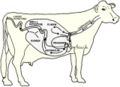 Cow rumen.jpeg 320 × 231; 21 KB
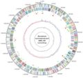 Genome real.jpg 700 × 658; 58 KB
 Bacteria.gif 250 × 250; 35 KB
 Chlamgrowthcycle.gif 483 × 464; 41 KB
 Fig12amats.gif 400 × 216; 29 KB
 Mature rb.gif 400 × 339; 45 KB
 Bordetella pertussis 01.jpg 829 × 764; 51 KB
 Bordetella pertussis 01.gif 829 × 764; 77 KB
 Ctepidum hotspring.jpg 598 × 896; 116 KB
 Bordetella pertussis 02.jpg 829 × 764; 51 KB
 1scipic3biofilms.jpg 263 × 350; 44 KB
 Helicobacterhepaticus2.gif 325 × 368; 40 KB
 Nature03661-f4.2.jpg 600 × 515; 71 KB
 File2.jpg 600 × 515; 71 KB
 Chlamydophila caviae.png 100 × 124; 16 KB
 Rick2-an.jpg 482 × 436; 90 KB
 U1fig10b.jpg 361 × 289; 32 KB
 Cationicpeptides(1).gif 587 × 326; 47 KB
 Shigella boydii 02.jpg 700 × 454; 15 KB
 Chlamidia.gif 300 × 200; 22 KB
 Propioni.jpg 750 × 1,002; 39 KB
 Propioni.jpeg 750 × 1,002; 39 KB
 Chlamydia-fig1.jpg 800 × 535; 82 KB
 Acnevulgaris.gif 557 × 350; 38 KB
 Adeh 2CPC .jpg 400 × 400; 36 KB
 Pacnesgenome.jpeg 600 × 600; 116 KB
 Ypseudo pillus.gif 100 × 104; 8 KB
 Yersinia pseudotuberculosis.GIF 346 × 132; 4 KB
 Glutamine.gif 832 × 828; 10 KB
 Glutaminee.gif 406 × 447; 4 KB
 Ehrlichia canis picture.jpg 300 × 200; 36 KB
 BIMM 120.JPG 444 × 213; 14 KB
 Cell96.gif 502 × 482; 80 KB
 APpic.png 278 × 277; 152 KB
 Picture of EM.jpg 500 × 325; 40 KB
 BIMM genome.JPG 401 × 401; 26 KB
 Genome.JPG 227 × 216; 9 KB
 Ima2ges.jpg 124 × 80; 5 KB
 Atlantica.jpg 484 × 44; 7 KB
 Genome pic.jpg 479 × 450; 36 KB
 Adbac.jpg 375 × 377; 31 KB
 Adbacteria.jpg 375 × 377; 34 KB
 Francois Thiaucourt.jpg 709 × 469; 13 KB
 Apek1 2.gif 220 × 220; 54 KB
 Bt6.JPG 829 × 345; 42 KB
 BorreliaII.GIF 153 × 104; 12 KB
 Thumb 1m.jpg 150 × 113; 3 KB
 Borrelia garinii.jpg 512 × 384; 19 KB
 Diag.jpg 521 × 426; 18 KB
 BorreliaII.gif 153 × 104; 12 KB
 Owen1b.jpg 328 × 446; 58 KB
 Owens4.gif 348 × 226; 31 KB
 Bt7.JPG 595 × 316; 40 KB
 E. coli genome.png 490 × 490; 129 KB
 BIMM.JPG 443 × 293; 12 KB
 Pic2-2.gif 386 × 1,033; 117 KB
 Leprosyvict.JPG 640 × 523; 58 KB
 1 1.png 600 × 600; 62 KB
 Number1 1.png 600 × 600; 62 KB
 Chart figure 1.jpg 140 × 97; 4 KB
 Chart figure 2.jpg 508 × 352; 26 KB
 C21c2.jpg 432 × 288; 71 KB
 Aquifex aeolicus budding.jpg 200 × 164; 4 KB
 030328 gutbugs.jpg 180 × 243; 10 KB
 Ralme.jpg 277 × 221; 53 KB
 Aquifex aeolicus.png 402 × 218; 63 KB
 US-Diphtheria.gif 770 × 512; 8 KB
 M.j..gif 225 × 225; 58 KB
 Water supply.jpg 700 × 460; 143 KB
 Methanococcus.jpg 300 × 200; 31 KB
 A02 bacteria full.jpg 518 × 656; 374 KB
 P01.jpg 240 × 162; 6 KB
 Hyphomonas.jpg 303 × 228; 11 KB
 Hyphomonasgenome.jpg 135 × 135; 4 KB
 Halorhodospira.gif 193 × 440; 71 KB
 Yeast.jpg 500 × 500; 160 KB
 240px-Streptococcus mutans 01.jpg 240 × 175; 12 KB
 240px-streptococcus mutans 01.jpg 240 × 175; 12 KB
 Picture 1.jpg 240 × 210; 35 KB
 OTSM.jpg 447 × 500; 77 KB
 Pg03c-1-.jpg 153 × 145; 10 KB
 Staphylococcus epidermidis lores.jpg 700 × 412; 55 KB
 Sporothrix 1.jpg 596 × 401; 46 KB
 Figure 1.JPG 935 × 1,209; 70 KB
 Periodontitis-1-.gif 226 × 227; 27 KB
 HCgramstain.jpg 150 × 100; 4 KB
 HCgramstain01.jpg 1,225 × 816; 82 KB
 HC02.JPG 640 × 400; 21 KB
 HC20C.JPG 640 × 400; 15 KB
 HC37C.JPG 640 × 400; 21 KB
 S.saprophyticus genome.png 600 × 600; 100 KB
 DennisKunkelMicroscopy,Inc.jpg 275 × 367; 61 KB
 C immitis structure.png 300 × 204; 123 KB
 26644C.jpg 275 × 324; 121 KB
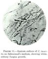 Coccidioides immitis on Sabouraud's medium.jpg 301 × 369; 45 KB
 Lactobacillus delbrueckii.jpg 72 × 48; 16 KB
 L delbrueckii subsp bulgaricus.jpg 72 × 48; 15 KB
 Stanford.jpg 311 × 149; 7 KB
 Meso 2.jpg 100 × 125; 10 KB
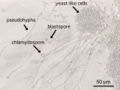 Image-C albicans en.jpg 640 × 480; 89 KB
 A.b.jpg 133 × 127; 3 KB
 Hmk.jpg 133 × 127; 3 KB
 96546F.jpg 349 × 275; 92 KB
 Chromosomes.jpg 480 × 320; 62 KB
 Aa microscope8.jpg 210 × 208; 10 KB
 DroppedImage 3.png 187 × 161; 28 KB
 Pdidemni cells.png 227 × 161; 50 KB
 Lpatella.png 241 × 161; 86 KB
 Alfalfa.JPG 300 × 202; 10 KB
 GenomeIloihiensis.jpg 400 × 566; 64 KB
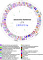
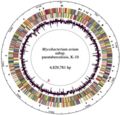 Map K10 Genome.jpg 440 × 421; 48 KB
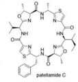 Patellamide.jpg 206 × 217; 7 KB
 Miniproch.jpg 62 × 68; 2 KB
 Amebiasis LifeCycle.gif 435 × 548; 28 KB
 Thermus Thermophilus.jpg 200 × 168; 23 KB
 Thermus.jpg 200 × 133; 59 KB


.gif)


