The History and Transmission of Treponema pallidum; Syphilis: Difference between revisions
No edit summary |
No edit summary |
||
| Line 9: | Line 9: | ||
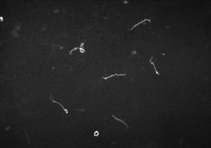
[[Image:TP.jpg|thumb|300px|right|Figure 1. Dark field microscopy depicts the presence of <i>Treponema pallidum<i/> the etiological agent responsible for Syphilis. The photo was taken in 1961 courtesy of the CDC's W.F. Schwartz. [http://http://phil.cdc.gov/phil/details.asp?pid=10179#modalIdString_CDCImage_0].]] | [[Image:TP.jpg|thumb|300px|right|Figure 1. Dark field microscopy depicts the presence of <i>Treponema pallidum<i/> the etiological agent responsible for Syphilis. The photo was taken in 1961 courtesy of the CDC's W.F. Schwartz. [http://http://phil.cdc.gov/phil/details.asp?pid=10179#modalIdString_CDCImage_0].]] | ||
<br>By: Marcus Townsend<br> | <br>By: Marcus Townsend<br> | ||
<br><br>The insertion code consists of: | |||
<br><b>Double brackets:</b> [[ | <br><b>Double brackets:</b> [[ | ||
<br><b>Filename:</b> PHIL_1181_lores.jpg | <br><b>Filename:</b> PHIL_1181_lores.jpg | ||
Revision as of 02:03, 29 April 2016
Classification
• Kingdom: Bacteria
• Phylum: Spirochaetae
• Class: Spirochaetes
• Order: Spirochaetales
• Family: Spirochaetacecae
• Genus: Treponema

By: Marcus Townsend
The insertion code consists of:
Double brackets: [[
Filename: PHIL_1181_lores.jpg
Thumbnail status: |thumb|
Pixel size: |300px|
Placement on page: |right|
Legend/credit: Electron micrograph of the Ebola Zaire virus. This was the first photo ever taken of the virus, on 10/13/1976. By Dr. F.A. Murphy, now at U.C. Davis, then at the CDC.
Closed double brackets: ]]
Other examples:
Bold
Italic
Subscript: H2O
Superscript: Fe3+
Introduce the topic of your paper. What is your research question? What experiments have addressed your question? Applications for medicine and/or environment?
Sample citations: [1]
[2]
A citation code consists of a hyperlinked reference within "ref" begin and end codes.
Description of Structure and Significance
Include some current research, with at least one figure showing data.
The bacteria Treponema pallidum is a bacteria prominently known for the cause of the chronic sexually transmitted disease syphilis. T. pallidum, also has relatives that are in a subspecies. These trepenomes include T. pallidum endemicum which causes Bejel, T. pallidum pertenue causes Yaws, and T. pallidum carateum causes Pinta. Phenotypically these four diseases appear similar along with their serology, but are very distinct based on their unique genotypes. T. pallidum genome is circular and is also very small in comparison to other prokaryotes as it contains roughly 1000 kilobase pairs in its genome. These distinct diseases are only found in humans. T. pallidum is classified as a spirochete, and its shape resembles that of a home telephone cord spiraling. Its structure is very thin and flexible. T. pallidum is considered a Gram-negative bacterium. Around its spiral shaped body, there is a cytoplasmic membrane which is enclosed by a second outer membrane. In between the cytoplasmic and outer membrane, there is a thin layer of peptidoglycan for stability. T. pallidum is a motile bacterium that possess endoflagella located in the periplasmic space between the two membranes, allowing the microbe to maneuver its environment, in what has been documented as in a corkscrew fashion. T. pallidum relies on chemotactic responses to be motile. The flagella contain 36 gene encoding proteins responsible for the structure and function of the flagella, along with two copies of the flagellar switch protein FliG. T. pallidum length ranges from 6-15µm in length and is 0.2 µm in width. T. pallidum is a small bacterium that requires a dark field microscopy to be visualized. Other techniques involved for visualizing this microbe include using a modified Steiner silver, the Dieterle stain or the Giemsa stain. This microbe is an obligate parasite as it needs a host to conduct its metabolic processes and life cycle; it is difficult to culture T. pallidum under in vitro conditions. T. pallidum thrives under low oxygen conditions from 10-20% levels, making it microaerophilic. It is also temperature dependent, as it prefers temperatures relative to the human body. T.pallidum is a primary pathogen because of its ability to compromise host’s immune system. As far as metabolic pathways are concerned, it does not contain a pathway to synthesize nucleotides, fatty acids, enzyme cofactors and most amino acids as well as lack genes to metabolize amino acids or fatty acids as a possible energy source. However, it does contain the necessities essential for glycolysis, but lacks genes responsible for steps that take place after glycolysis. T. pallidum lacks genes that would allow products from glycolysis to enter the TCA cycle, and eventually the electron transport chain. Lastly, it lacks genes that allows oxidative phosphorylation pathways to occur. Due to T. pallidum missing integral components for the TCA cycle and the ETC, its one dimensionality based solely on glucose metabolism via glycolysis, it cannot optimize its energy production. T. pallidum also has a specific set of genes that code for 18 unique ATP-binding cassette transporters with specificity for certain molecules. The limited pathway ability of T. pallidum can be attributed to the small genome it possess. It is heavily reliant upon the conditions the host supplies it with. These factors make it difficult for scientist to readily culture T. pallidum because of the ideal conditions it requires and the limited pathways it has.
Every point of information REQUIRES CITATION using the citation tool shown above.
History and Discovery
Include some current research, with at least one figure showing data.
Transmission and Symptoms
Include some current research, with at least one figure showing data.

Section 4
Conclusion
References
Authored for BIOL 238 Microbiology, taught by Joan Slonczewski, 2016, Kenyon College.
