|
|
| Line 39: |
Line 39: |
| <br>Include some current research, with at least one figure showing data.<br> | | <br>Include some current research, with at least one figure showing data.<br> |
|
| |
|
| [[Image:h pylori data.jpg|thumb|450px|left|Fig. 2. Scanning electron micrograph of <i>Lactobacillus bulgaricus</i>. Taken from Wang et al., 2004.]] | | [[Image:h pylori data.jpg|thumb|450px|left|Fig. 7. Scanning electron micrograph of <i>Lactobacillus bulgaricus</i>. Taken from Wang et al., 2004.]] |
|
| |
|
|
| |
|
Revision as of 09:51, 17 April 2010
Introduction
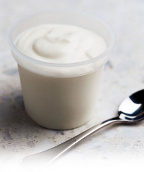
Fig. 1. Yogurt as often seen and consumed.
At right is a sample image insertion. It works for any image uploaded anywhere to MicrobeWiki. The insertion code consists of:
Double brackets: [[
Filename: PHIL_1181_lores.jpg
Thumbnail status: |thumb|
Pixel size: |300px|
Placement on page: |right|
Legend/credit: Electron micrograph of the Ebola Zaire virus. This was the first photo ever taken of the virus, on 10/13/1976. By Dr. F.A. Murphy, now at U.C. Davis, then at the CDC.
Closed double brackets: ]]
Other examples:
Bold
Italic
Subscript: H2O
Superscript: Fe3+
Biochemistry of Yogurt Production

Fig. 2. Overview of biochemical processes in yogurt production. Courtesy of The Food and Agriculture Organization of the United Nations.
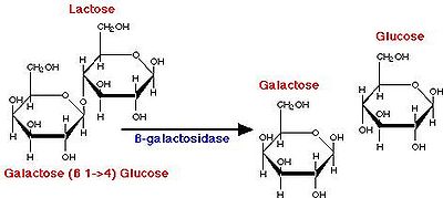
Fig. 3. Lactose catabolism into glucose and galactose. Courtesy of Thomas M. Terry at the University of Hamburg.
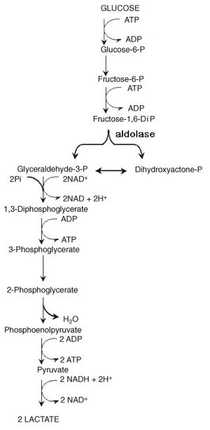
Fig. 4. Glycolysis and homolactic fermentation. Courtesy of Dr. Todar's Online Textbook of Bacteriology.
Introduce the topic of your paper. What microorganisms are of interest? Habitat? Applications for medicine and/or environment?
Yogurt Production
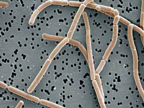
Fig. 5. Scanning electron micrograph of
Lactobacillus bulgaricus. Courtesy of The Microscopy Facility at Utah State University.

Fig. 6. Scanning electron micrograph of
Streptococcus thermophilus. Courtesy of Dennis Kunkel Microscopy, Inc.
Benefits of Yogurt
Include some current research, with at least one figure showing data.
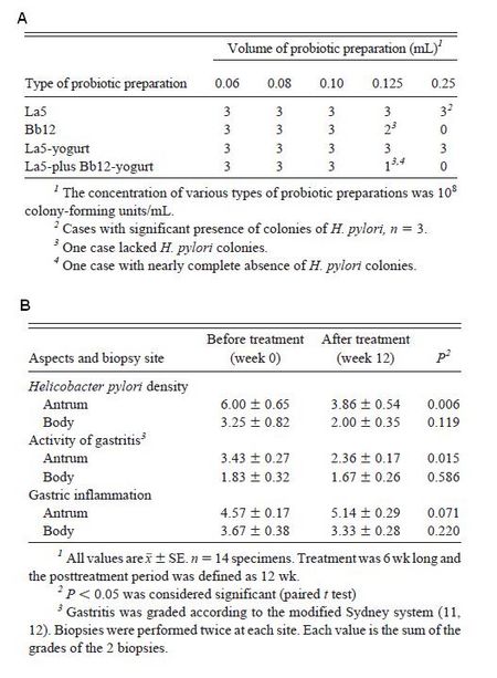
Fig. 7. Scanning electron micrograph of
Lactobacillus bulgaricus. Taken from Wang et al., 2004.
Probiotics
Include some current research, with at least one figure showing data.
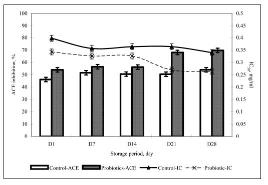
Fig. 2. Scanning electron micrograph of
Lactobacillus bulgaricus. Taken from Donkor et al., 2005.
Lactobacillus casei
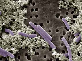
Fig. 2. Scanning electron micrograph of
Lactobacillus casei. Courtesy of The Microscopy Facility at Utah State University.
Lactobacillus acidophilus
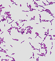
Fig. 2. Scanning electron micrograph of
Lactobacillus acidophilus. Courtesy of Dr. Todar's Online Textbook of Bacteriology.
Bifidobacterium species

Fig. 2. Scanning electron micrograph of
Bifidobacterium. Courtesy of Dr. Sandy Smith, Dept. of Food Science, University of Guelph, Canada.
Improving Yogurt
Current Problems
Include some current research, with at least one figure showing data.
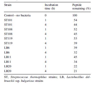
Fig. 2. Scanning electron micrograph of
Lactobacillus bulgaricus. Taken from Paul and Somkuti, 2010.
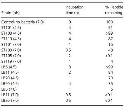
Fig. 2. Scanning electron micrograph of
Lactobacillus bulgaricus. Taken from Paul and Somkuti, 2009.
Improving functionality of Yogurt
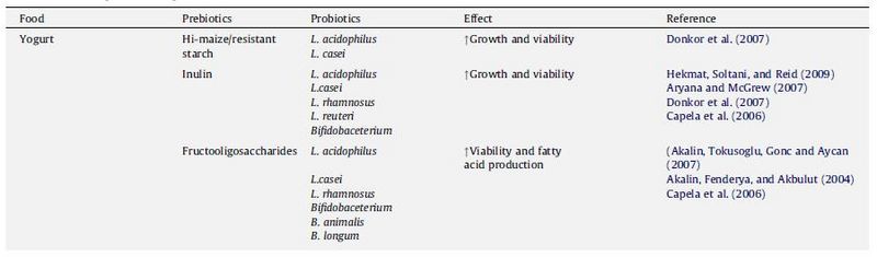
Fig. 2. Scanning electron micrograph of
Lactobacillus bulgaricus. Taken from Ranadheera, Baines, & Adams, 2009.
A "Superior" Yogurt
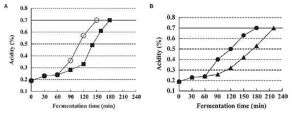
Fig. 2. Scanning electron micrograph of
Lactobacillus bulgaricus. Taken from Ranadheera, Baines, & Adams, 2009.
Fig. 2. Scanning electron micrograph of
Lactobacillus bulgaricus. Taken from Ranadheera, Baines, & Adams, 2009.

Fig. 2. Scanning electron micrograph of
Lactobacillus bulgaricus. Taken from Ranadheera, Baines, & Adams, 2009.
Conclusion
Overall text length at least 3,000 words, with at least 3 figures.
References
[Sample reference] Takai, K., Sugai, A., Itoh, T., and Horikoshi, K. "Palaeococcus ferrophilus gen. nov., sp. nov., a barophilic, hyperthermophilic archaeon from a deep-sea hydrothermal vent chimney". International Journal of Systematic and Evolutionary Microbiology. 2000. Volume 50. p. 489-500.
Edited by student of Joan Slonczewski for BIOL 238 Microbiology, 2010, Kenyon College.


