Trichoplax
Classification
Domain - Eukarya; Phylum - Placozoa; Family - Trichoplacidae
Species
Genus species: Trichoplax adhaerens
|
NCBI: Taxonomy [1] |
Description and Significance
Trichoplax are the smallest multicellular animal known in science. Their diameter is only 1-2mm and they appear as flat, disc-shaped, and have no body symmetry. They are found in tropical and sub-tropical environments. Trichoplax Adhaerens, the only concretely defined species within the genus, has the smallest DNA sequence discovered within an animal. Trichoplax adhaerens is viewed as a potential biological model organism as it moves via locomotion and coordinates without full-fledged muscle and nerve tissue. Scientists often think that Trichoplax allows us to imagine how the earliest animals were organized and compartmentalized before complex structures like mouths and nerves. Thus, Trichoplax can serve to be a indicator of important evolutionary processes regarding eukaryotes, allowing scientists to further understand the development of animals.
Genome Structure
Other interesting features? What is known about its sequence?
Trichoplax's genome is the smallest genome of any animal measured. it consists of six haploid chromosomes. Morphology of the chromosome is unclear. Inside the chromosomes, there are 11,584 genes which are encoded by ~98,000,0000 base pairs.
Cell Structure, Metabolism and Life Cycle
Interesting features of cell structure; how it gains energy; what important molecules it produces.
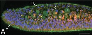
Trichoplax only has 6 cell types. 4 of the 6 are incorporated into the epithelium that encloses it. The epithelium divides into two parts: the upper and lower epithelium. The upper epithelium is thin and the lower is thicker. As cells transition closer to the lower epithelium, cells become progressively more dense. This creates a pseudostratified arrangement. Between the upper and lower epithelium are fiber cells. These fiber cells contact each other by branching and also allow communication between all other cells. Typical gland cells are also present. They are present around the circumference in a ring-like manner as well as sparsely in the organism. They contain neuropeptides which express neurosecretory proteins. There are also crystal cells, containing birefringent crystals, are distributed at near the edge of the animal.
There are two main cells in epithelium that aid in the feeding process. The prevalent cell type within the epithelium are columnar cells. They contain a single cilium and multiple microvilli. The single cilium is what allows the cell glide via locomotion. Gliding occurs until algae becomes present, where the Trichoplax halts to feed. This is where the other cell type, non-cilliated lipophil, comes into play. Non-ciliated lipohill cells are composed of large granules and has an even bigger granule proximal on the surface of the upper epithelium. This is where the the algae is ingested through the ventral (upper epithelium) granule. Neurosecretery cells may also have a hand in directing the cell while eating due to the neuropeptides. This is similar to animals that are far more complex.
Ecology and Pathogenesis
Habitat; symbiosis; biogeochemical significance; contributions to environment.
If relevant, how does this organism cause disease? Human, animal, plant hosts? Virulence factors, as well as patient symptoms.
Trichoplax live in aquatic environments that are rich in seawater. It has rarely been observed in the wild, leaving many questions regarding its benefit to the environment.
References
Author
Page authored by Mark Peck II, student of Dr. Bradley Tolar at UNC Wilmington.

