Viral Replication and Ebola
Introduction
The ebola virus genus which is responsible for the cause of Ebola Virus Disease (EVD), a hemorrhagic fever virus in humans. The U.S. Centers for Disease Control and Prevention (CDC) has classified Ebola as a Category A select agent. The genome of Ebolaviruses (EBOV) consists of non-segmented, single-stranded RNA structures. EBOV belongs to the virus family Filoviridae which it shares with Marburgvirus and Cuevavirus.
(Chart) Identified Species of Ebola Virus (EBOV) Zaire ebolavirus (ZEBOV) Bundibugyo ebolavirus (BDBV) Sudan ebolavirus (SUDV) Taï Forest ebolavirus (TAFV) Reston ebolavirus (RESTV) Bombali
Along with SUDV, ZEBOV is one of the most common outbreak-causing pathogens known for its inducement of the largest outbreak in West African history. Without treatment, ZEBOV claims a fatality rate of up to 90%, making it the most lethal strain of EVD.(5) Although the fatality rates vary from case to case, the current fatality rate of EVD has decreased significantly due to heightened awareness of its transmission mechanisms and effective treatments. Currently, the average fatality rate of EVD patients who receive treatment is about 50%.
The onset of symptoms typically occur between the incubation period of 2-21 days after exposure to Ebola.
(Chart) Early Symptoms of EBOV Advanced Symptoms of EBOV Vomiting Fever Headache Abdominal pain Diarrhea Bleeding from all orifices Multi-organ failure Hypovolemic shock Coffee ground emesis
Jeanne Kantungo, a 38 year-old mother and Ebola survivor from The Democratic Republic of Congo, shares her experience with the exigency of Ebola coupled with its symptoms. In Kantugo’s interview with The International Rescue Committee, she states:
“It started with headaches...then blood all over my body. I was in the Ebola Treatment Center for two months. I felt so lonely. Doctors and nurses reassured me that I would recover and go home. Kids were sad when they saw me going. They thought I would die.”
Transmission of EBOV occurs through direct contact of contaminated body fluids with mucous membranes and open wounds as they are direct pathways to the bloodstream. Typical contaminated body fluids to avoid are: blood, saliva, sweat, tears, mucus, vomit, feces, and urine (2). In addition, Ebola can be atypically transmitted via aerosols, infectious droplets released into the air. This mode of transmission has been documented to allow the virus to spread in medical facilities when patients undergo certain medical procedures ( ). Furthermore, Ebola survivors can carry the viral RNA in bodily fluids such as breast milk and semen for up to 200 days. According to ____ approximately 69.2% of patients tested positive after 3 to 6 months…
(Graph)
While the origins of Ebola are indefinite, the zoonotic disease has led scientists to believe that the initial hosts of the virus are fruit bats of the Pteropodidae family, also known as “old world bats” (Figure 3). These megabats, which are found in various parts of Africa, are considered to be a natural reservoir of the pathogen. Other animals such as apes, forest antelopes, and porcupines are known to have been infected. Ebola was most likely transmitted to humans via hunters who came in direct contact with the infected animals or sold them for consumption.
The first Ebola case occurred during the year 1976 in Zaire, which is now known as the Democratic Republic of Congo, located near the Ebola River(). Following the initial case was an outbreak in Sudan which resulted in a mortality rate of roughly 53%. Up until the year 2000, within sub-saharan Africa, Ebola primarily affected Central and East African countries. 2014 marked the largest Ebola outbreak in African history, initially spreading between Guinea, Liberia, and Sierra Leone. The outbreak resulted in the infection and death of tens of thousands of people. Zaire ebolavirus, the most acquired strain in the West African outbreak, expanded further westward, reaching the countries of Nigeria, Mali, and Senegal. Between 1990 and 2016 Ebola cases were found within 6 countries outside of Africa: United States, United Kingdom, Spain, Russia, and Italy. The cause of these cases mostly came from healthcare workers who were treating patients at Ebola treatment centers in Africa and lab accidents. Ebola cases further arose from infected airline passengers and animals, specifically pigs and macaques from The Philippines. Infected macaques which were imported to a primate holding facility in the United States, were responsible for the well known cases from Reston, Virginia. The very last major Ebola outbreak which occurred in Uganda during the fall of 2022, ended months later in early 2023. During this outbreak 164 total cases along with 77 deaths were reported.
New Section

By [Author Name]
At right is a sample image insertion. It works for any image uploaded anywhere to MicrobeWiki.
The insertion code consists of:
Double brackets: [[
Filename: PHIL_1181_lores.jpg
Thumbnail status: |thumb|
Pixel size: |300px|
Placement on page: |right|
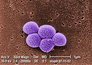
Legend/credit: Magnified 20,000X, this colorized scanning electron micrograph (SEM) depicts a grouping of methicillin resistant Staphylococcus aureus (MRSA) bacteria. Photo credit: CDC. Every image requires a link to the source.
Closed double brackets: ]]
Other examples:
Bold
Italic
Subscript: H2O
Superscript: Fe3+
Sample citations: [1]
[2]
A citation code consists of a hyperlinked reference within "ref" begin and end codes.
To repeat the citation for other statements, the reference needs to have a names: "<ref name=aa>"
The repeated citation works like this, with a forward slash.[1]
