Virulence of Pseudomonas savastanoi: Difference between revisions
| Line 4: | Line 4: | ||
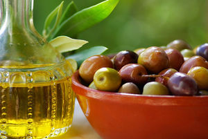
[[Image:Olive-oil-and-mature-olives.-.jpg|thumb|300px|right|Promoting olive crops has been of interest due to the proven health benefits of the Mediterranean diet, which includes olives and olive oil. Source: http://www.walksofitaly.com]] | [[Image:Olive-oil-and-mature-olives.-.jpg|thumb|300px|right|Promoting olive crops has been of interest due to the proven health benefits of the Mediterranean diet, which includes olives and olive oil. Source: http://www.walksofitaly.com]] | ||
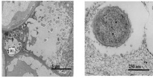
[[Image:Hildy pic 1.jpg|thumb|300px|right|Visualization of outer membrane vesicles, which help facilitate P. savastanoi pathogenicity. (left)Knot tissue invaded by P. savastanoi microbes. (right) Release of outer membrane vesicles forming from host cell. Modified from Pérez-Martínez et al. 2010]] | [[Image:Hildy pic 1.jpg|thumb|300px|right|Visualization of outer membrane vesicles, which help facilitate P. savastanoi pathogenicity. (left)Knot tissue invaded by P. savastanoi microbes. (right) Release of outer membrane vesicles forming from host cell. Modified from Pérez-Martínez et al. 2010]] | ||
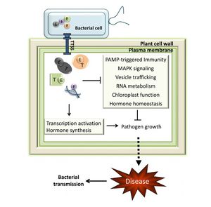
[[Image:TTSS.jpg|thumb|300px|right|General mechanism of Type III Secretion System (T3SS) in plant pathogens. (Melotto and Kunkel 2013)]] | |||
Revision as of 01:27, 25 April 2013
Introduction


By: Hildy Joseph, Kenyon 2013
At right is a sample image insertion. It works for any image uploaded anywhere to MicrobeWiki. The insertion code consists of:
Double brackets: [[
Filename: PHIL_1181_lores.jpg
Thumbnail status: |thumb|
Pixel size: |300px|
Placement on page: |right|
Legend/credit: Electron micrograph of the Ebola Zaire virus. This was the first photo ever taken of the virus, on 10/13/1976. By Dr. F.A. Murphy, now at U.C. Davis, then at the CDC.
Closed double brackets: ]]
Other examples:
Bold
Italic
Subscript: H2O
Superscript: Fe3+
Introduce the topic of your paper. What microorganisms are of interest? Habitat? Applications for medicine and/or environment?
Section 1
Include some current research, with at least one figure showing data.
Section 2
Include some current research, with at least one figure showing data.
Section 3
Include some current research, with at least one figure showing data.
Conclusion
Overall text length at least 3,000 words, with at least 3 figures.
References
Edited by student of Joan Slonczewski for BIOL 238 Microbiology, 2011, Kenyon College.


