Yersinia pestis, the History of the Plague and Adaptation to Animal Host: Difference between revisions
| Line 28: | Line 28: | ||
==Discovery== | ==Discovery== | ||
Include some current research, with at least one figure showing data.<br> | Include some current research, with at least one figure showing data.<br> | ||
<br> | <br> | ||
Alexandre Yersin first discovered <i>Yersinia pestis</i> in 1894 at the Pasteur Institute, while a plague occurred in Hong Kong, cause by this <i>Y. pestis</i> bacterium. Yersin was the first to link the plague to the <i>Y. pestis</i> bacterium and therefore the original name of the bacterium, <i>Pasteurella pestis</i>, was changed to the modern name, <i>Yersinia pestis</i>. This outbreak in China, which began in 1855, was said to have killed over 12 million people in both India and China, and was not considered over until 1959. There have been three major outbreaks of plague caused by these bacteria, the plague of Justinian, the Black Death, and the third plague pandemic. <br> | |||
==History== | ==History== | ||
Revision as of 23:26, 21 April 2015
Classification
• Kingdom: Eubacteria
• Phylum: Proteobacteria
• Class: Gammaproteobacteria
• Order: Enterobacteriales
• Family: Enterobacteriaceae
• Genus: Yersinia
Structure and Significance

By [Blake Calcei]
At right is a sample image insertion. It works for any image uploaded anywhere to MicrobeWiki. The insertion code consists of:
Double brackets: [[
Filename: PHIL_1181_lores.jpg
Thumbnail status: |thumb|
Pixel size: |300px|
Placement on page: |right|
Legend/credit: Electron micrograph of the Ebola Zaire virus. This was the first photo ever taken of the virus, on 10/13/1976. By Dr. F.A. Murphy, now at U.C. Davis, then at the CDC.
Closed double brackets: ]]
Other examples:
Bold
Italic
Subscript: H2O
Superscript: Fe3+
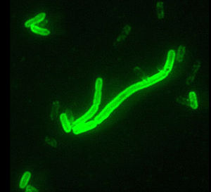
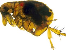
Yersinia pestis, is most notable for the its devastating role in plagues of the past. These bacteria are Gamaproteobacteria that are Gram negative with a coccobacillus shape and are facultative anaerobes. This bacterium causes infection in humans and other non-human animals. These bacteria can be found in animal or insect blood, and this blood is required for the bacteria to survive. The most common hosts of these bacteria include, insects (fleas), rodents and humans. The most notable carrier of this bacteria is the Oriental Rat flea, which contains bacteria in its blood but is unaffected by the bacteria. These fleas then infect rats, which eventually pass the bacteria to humans and other non-human animals. The three main infections caused by this bacterium include: pneumonic, septicemic and bubonic plagues. These diseases are also commonly found in highly populated areas that have poor sanitation habits therefore increasing the rat population and making the location prime for Y. pestis infections. The progression from fleas to rats and the widespread populations of both of these organisms has allowed these bacteria to be exposed to a wide variety of environmental conditions allowing it to adapt to multiple strains that are very resilient. The regulatory systems involved in the virulence of these bacteria have been found to be quite complex posing a large problem for antibiotics.
Discovery
Include some current research, with at least one figure showing data.
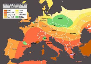
Alexandre Yersin first discovered Yersinia pestis in 1894 at the Pasteur Institute, while a plague occurred in Hong Kong, cause by this Y. pestis bacterium. Yersin was the first to link the plague to the Y. pestis bacterium and therefore the original name of the bacterium, Pasteurella pestis, was changed to the modern name, Yersinia pestis. This outbreak in China, which began in 1855, was said to have killed over 12 million people in both India and China, and was not considered over until 1959. There have been three major outbreaks of plague caused by these bacteria, the plague of Justinian, the Black Death, and the third plague pandemic.
History
Include some current research, with at least one figure showing data.

Yersinia pestis Adaptation to Animal Host
Include some current research, with at least one figure showing data.

Methods for Prevention and Vaccines
Include some current research, with at least one figure showing data.
Conclusion
Include some current research, with at least one figure showing data.
