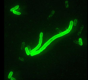
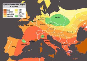
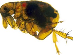
Yersinia pestis, the History of the Plague and Adaptation to Animal Host
Structure and Significance

By [Blake Calcei]
At right is a sample image insertion. It works for any image uploaded anywhere to MicrobeWiki. The insertion code consists of:
Double brackets: [[
Filename: PHIL_1181_lores.jpg
Thumbnail status: |thumb|
Pixel size: |300px|
Placement on page: |right|
Legend/credit: Electron micrograph of the Ebola Zaire virus. This was the first photo ever taken of the virus, on 10/13/1976. By Dr. F.A. Murphy, now at U.C. Davis, then at the CDC.
Closed double brackets: ]]
Other examples:
Bold
Italic
Subscript: H2O
Superscript: Fe3+
Introduce the topic of your paper. What microorganisms are of interest? Habitat? Applications for medicine and/or environment?
Discovery
Include some current research, with at least one figure showing data.
History
Include some current research, with at least one figure showing data.

Yersinia pestis Adaptation to Animal Host
Include some current research, with at least one figure showing data.

Methods for Prevention and Vaccines
Include some current research, with at least one figure showing data.
Conclusion
Include some current research, with at least one figure showing data.
