Abutilon mosaic virus
By: Deveren Manley II
Introduction
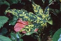
Abutilon mosaic virus (AbMV) is a plant virus that infects evergreen upright shrubs, Abutilon striatum. The first recognized infection was in 1868, in which one shrub in a shipment of A. striatum brought to England had the signature bright yellow mottling symptom of the pathogen. Abutilon mosaic virus is a part of the Geminivirus/Geminiviridae family and Begomovirus genus. Viruses in this genus have a insect vector transmission cycle. The vector for this virus is the silverleaf whitefly, scientifically known as Bemisia tabaci. The prevalence of the whitefly worldwide allows the virus to be just as widely prevalent. There are 3 strains of the AbMV: Abutilon mosaic A: West Indies virus, Abutilon mosaic B: Brazil virus, and Abutilon mosaic Hawaii virus. All strains are appreciated by botanists and plant lovers because they add decorative color to their plant host with little negative effect to life cycle and health. [1]
Structure
AbMV characteristics from the Geminivirus family
Abutilon mosaic virus infects weeds, crops, and ornamentals. They are mostly harmless to hosts unlike relatives in the family that can cause serious damage to crop land. Abutilon mosaic virus has a circular, single-stranded DNA (ssDNA) structure that readily recombines and mutates. Like other geminiviruses, they are unable to replicate through methylation, and take advantage of host cellular mechanisms to accomplish this task.[2] Their ability to acquire novel genes allows them to adapt quickly to the vastly different hosts and environments they may encounter across the world. [3]
AbMV characteristics from Begomovirus Genus
Abutilon mosaic virus has 321 peers in Begomoviruses Genus. The DNA of these species are packaged into a nucleocapsid that is 38 nanometers (nm) long and 15–22 nm in diameter. Each capsid is made of 22 capsomeres.[4] The single stranded DNA inside the capsid are not enveloped, adopting icosahedral symmetry.
Begomoviruses have a bipartite genome, in which both segments of DNA are required for successful infection of a host cell. The DNA in the first segment encodes for replication, transport, regulatory, and coat proteins. Specifically they control how genes are expressed, ability to evade or defeat host defenses, capsid formation, and transmission to vector. This segment of DNA is shared in all geminiviruses. The second segment of DNA encodes for viral movement and symptom expression. [5]
Transmission
AbMV life cycle: plant to whitefly
Bemisia tabaci, also known as the silverleaf whitefly, is the vector of Abutilon mosaic virus. The virus is transmitted to whiteflies through their digestive system. The virus enters while it feeds on an infected plant. The longer the whitefly enjoys its plant meal, the more likely it is to contract the virus. Once inside the whitefly, AbMV inhabits the salivary glands, midgut, and filter chamber. After fully establishing itself inside the whitefly, AbMV does not reproduce inside it. Despite the lack of reproduction, the whiteflies remain infected for life, including molts, similar to Herpes simplex virus in humans. In contrast to Herpes, the virus cannot be passed from an infected whitefly onto its offspring. Overtime, as the infected whitefly becomes older, the virus becomes attenuated losing its ability to continue the transmission cycle into other vectors. [6]
AbMV life cycle: whitefly to plant

When whiteflies first contract AbMV there is a 12 hour window that must elapse before it can transmit the virus to plant hosts. After this latency period, the infected adult whiteflies begin to transmit the virus when they feed on the next Abutilon species.[7] Speaking from an evolutionary standpoint, whiteflies are a sound vector selection because they feed on many types of plant species giving the virus the best opportunity to be transmitted to many plants. Once inside the plant, AbMV virus molecules congregate in the nuclei of bundle area cells.[8] After successful transmission, the virus will likely inhabit the plant for the rest of its life, just as the whitefly. In a natural environment, Abutilon species continue the transmission cycle when the next whitefly feeds on its leaves.
Non-organismal Mode of Transmission
Present day, AbMV is most abundantly transmitted through grafting across Abutilon striatum populations. The aesthetic color patterns caused by the virus on host leaves generate consumer interest. Gardeners and botanists intentionally spread the virus to meet consumer demand. Outside of the spread of AbMV, gardeners also use grafting to:
• Get immature plants to produce fruit
• Pass on beneficial genes
• Repair damage to trees stripped of bark
• Conduct plant research
The purpose of the technique is to create contact between the vascular systems of two plants establishing a pathway of gene flow and nutrient-sharing. Grafting allows plants to share cell nuclei, DNA, growth stimuli and chloroplasts.[9] When trying to directly transmit AbMV using this method, the best connection is between the rootstock of an infected plant and the scion of the non-infected plant.[10] The genetic similarity between the two plants are important to the formation of a connection between them. The closer they are in species to one another, the more likely they will grow together.
To graft plants together, the desired areas of both plants are peel back to expose their vascular layers using a knife. The plants are bound together using grafting tape or wax where their vascular system is exposed. (The adhesives also reduce nutrient loss due to vascular system exposure.) The two vascular areas remain compressed together over the span of weeks until the grafting process has been completed. When the two plants have been united scar tissue will have formed on the grafted area.[11] Once successfully grafted the plants will grow together and the AbMV infection should be present in both plants. (Researchers have used grafting to study transmission cycles of other viruses well. They created a Viral Index based on their findings using this method of transmission. They first graft a plant suspected to be a non-symptomatic viral carrier to a plant that is very susceptible to the suspected virus. If the susceptible plant becomes infected they know that the initial plant is a carrier, and is added to the Viral Index.[12])
Viral Vectors

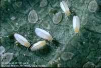
Bemisia tabaci was first identified in the United States 1896, not long after the first infection of AbMV in England. This species of whitefly are extremely small at about 1.5 mm long.[13] They have a yellow torso with white wings. Their environmental preference tends to include the warm climate associated with the tropical, subtropical, and sometimes temperate habitats. This prevalence in warmer climates is not by chance as cold temperatures kill them.
Whiteflies suck the nutrients out of plants, which is why they are mostly found on the bottom of leaves. This method of nutrition is harmful to plants because it leads to poor growth, shedding, and lower crop yields. Outside of stealing nutrients from plants, it also corrupts plant life because it normally is a host for multiple diseases. Since its introduction to the United States, pathogen-carrying whiteflies have cost the agricultural industries in Texas and California over $100 million worth of crops. Researchers have not been able to figure out how it has been able to evade its natural predators which include lady beetles and several species of wasp. To improve crop yields, pesticides have been created to kill whiteflies, but these treatments have led to resistant strains.
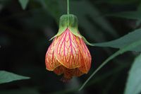
Abutilon striatum are versatile plants that can be found in a number different areas. It naturally finds its home in South American countries such as Paraguay, Uruguay, Argentina, and Brazil, where there is a warm climate. The flower is mainly known for its colorful structure and is a popular ornamental house plant kept in pots anywhere in homes. There are no strict requirements for sun exposure but this does have a small effect on plant health in terms of vibrancy. They normally grow to be 5 meters tall and 2 meters with leaves that grow to be anywhere between 5-15 cm long. The petals of the shrub grow to be around 2-4 centimeters long. Their flowers are normally yellow or orange and have red veins. They begin to bloom in the spring during the month of April all the way through the summer until September. In countries with regular warm weather the bloom lasts longer such as where it naturally grows in South America. The shrubs do attract bees and other pollinators alike who the plants use to spread their pollen. Abutilon striatum can be eaten by humans. They are able to be cooked but do not have to be. They have a sweet taste to them.
AbMV Physiological Impacts on Abutilon striatum

Once a plant is infected with the virus it is likely infected for the rest of its life. Although the plant remains infected the amount of damage the disease causes is minimal. It is not a death sentence. Plants will still be able to bloom, growth, pollenate, etc. There may be some stunting of growth, dehydration, and decrease in the production of photosynthesis. The symptoms a plant experiences are not distinctly defined. Symptoms displayed often depend on the species of Abutilon infected and the strain of the virus encountered. The Hawaii strain of the virus causes crinkling and mottling in the leaves of the plants it infects. It does not cause the desired yellow mosaic pattern coveted by most botanist. Another key difference in the Hawaii strain from the other strains is that it is not transmissible through the whitefly vector.
Much of what is known about the Abutilon mosaic virus physiology has been obtained through research. There have been research done to make sure it has fulfilled all 4 of Robert Koch’s postulates. The second postulate of the ability to obtain in pure culture was hard to fulfill when first discovered because the disease is in low concentration in the vascular system of the plant. To satisfy the first two postulates the genome of AbMV was sequenced from a disease plant and then compared the amount of similarity in DNA between a host plant and uninfected plant. The process of DNA comparison for similarity is called a DNA hybridization test. Then researchers used an inoculation method in which they rubbed the disease onto the vascular system on a healthy plant which is called mechanical inoculation. After the disease was successfully spread then it was re-isolated and tested using the same methods used to satisfy the first and second postulate. [14]
At a molecular level the virus only replicates in the phloem of the plant.[15] The virus has the ability to take the red color out of the veins of Abutilon and with normally form the coveted color spots in between the veins. The effects of the disease are strongly linked to photosynthetic output of the plant. This means the more light the plant receives the more the plant will experience symptoms. The virus also causes carbohydrate accumulation in the leaves as well. Things such as starches, sucrose, and hexose are associated with infection. [16]
Researchers have not developed a cure for this virus because it does little damage to the plant host. It is recommended that plants infected with the virus whose growth is severely stunted should be replaced. Before you completely uproot an infected plant you can also try to compensate for the constraints placed on the plants by the virus by taking great care of the plant. Making sure it receives high levels of sun, the right amount of water, and rooted in the correct soil. All these little things can improve the health of an ailing plant. There also has been some success in cutting off symptomatic leaves and allowing them to regrow. This method is not 100% guarantee to cure the virus but does provide the plant with an opportunity to get rid of the virus.
The carbohydrates or sugars buildup in the leaves as a product of photosynthesis. Under normal circumstances these carbohydrates a removed from the leaves by other mechanisms of the plant to distributed to the rest of the organism. Abutilon mosaic virus interferes with this process not allowing them to be transported. This interaction with sugar production is why the virus is most active after periods of high photosynthesis. The more sugars made the more it blocks. This buildup of sugar degrades the leaves photosynthetic systems which is why the leaves develop the mosaic pattern. The vibrant green color in leaves is a product of healthy chlorophyll conducting photosynthesis. An interruption in this process cause the chlorophyll to lose this pigment. High carbohydrate contents in the phloem alone.
When you consider, Abutilon mosaic virus’ attack on the phloem or vascular system of plants makes that the most common method of transmission is through grafting. The physical connection between the two plants’ vascular system gives the virus a direct pathway to travel to the uninfected plant’s photosynthetic organelles and cause the same carbohydrate buildup. This buildup of carbohydrates also explains why some plants experience a stunting of growth, withering, and crinkling. The carbohydrate buildup can be thought of as a blood clot in humans that causes swelling. If the clot goes untreated there is loss of function, color change due to being oxygen deprived among other symptoms. Plants experience the same thing. Crinkling, mosaic patterns, etc. are the plants form of clotting the area. The attenuation the virus experiences across time is one factor that explains why a range of symptoms is seen in infection. For example, passing on an old attenuated virus is a healthy plant in a shaded environment likely will not experience a large degree of symptoms.
Current Research: ABMV promoted carbohydrate buildup's effect on plant photosynthetic systems Lohaus et al.
A research team conducted a study in Australia at the National University in 2008, conducted a 3-month study analyzing Abutilon mosaic virus’ stages of symptom development through the scientific lens of localized carbohydrate progressive buildup and impaired photosynthetic output in Abutilon species plants. Researchers collected more than one sample from each plant in the study. One sample was taken from a portion of the infected plant’s leaf displaying the symptomatic mosaic pattern. The other sample was taken from an asymptomatic green portion of the same leaf. This data was used in tandem as a measure of symptom intensity. Symptom intensities were analyzed across a population of Abutilon whom ranged from recent infection to plants who have had the virus for an extended period. Most of these long-term carriers of the virus referred in the study as “mature” had a weakened form of the virus as it had evolved away from its most virulent wild type form.
These researchers were interested in studying this Abutilon mosaic virus- Abutilon striatum virus-host infection development to gain a better understanding of viral symbiotic mechanisms in a setting that did not require a large amount of safety precautions. Specifically, in terms of plant physiology the results of this study directly relate to the degree impaired photosynthetic output impacts plant growth.
Some plants for this study were kept in potting soil in a glass house allowing them to receive natural sunlight. Other plants were stored in an growth room the controlled for temperature during the day and night. The temperature during the day was 25⁰C and the temperature at night was 20⁰C[17]. All plants were systematically infected using the same grafting methods. Mature leaves and young leaves were selected based on expression of yellow mosaic pattern symptoms between the leaf veins. Carbohydrate samples were hole punched semi-randomly from symptomatic and asymptomatic portions of the leaf. Sample leaf disc were 4mm in diameter, with 16 samples coming from one leaf. Samples were collected at the end of the day and at the end of the night. After the sample was taken it was then placed in liquid nitrogen and stored at -70⁰C until it was time for further analysis. To begin the content analysis, each disc was grinded into small particles on -70⁰C media. The sample was then treated with chloroform and methanol and mixed into an aqueous mixture. After the mixture was formed it was kept on ice for 30 minutes. After the time elapsed, the extracted the sample with a total of 6 mL of water. The liquid treatments were then evaporated off using a rotary evaporator so only the solid phase was left of the sample. The solid phase was then treated with another 2 mL of water and stored at -80⁰C[18]. Carbohydrates were detected from these samples based on their acid/base properties via anionic exchange using sodium hydroxide. The content of these samples were measured in milli-moles.
Photosynthetic expression was expressed through “chlorophyll fluorescence quenching”[19]. This a technique that uses technology to take pictures of leaves at different light intensities. The camera is can detect induction caused by photosynthesis from changes in the air surrounding the leaf plant. Samples for this testing were also taken from the glasshouse and growth room. Each desired subject leaf was enclosed in a leaf case that darkened the surrounding environment. This served as a pre-treatment method to control for any photosynthetic outputs being conducted by the plant before the test. The quenching technology stimulates photosynthesis in the plant using a red LED light. The technology flashes the red LED light in 2 second intervals at various energy of light. The intensity of light is known by the machine. The technology then using reading from the images and calculations allows the researchers to see photosynthetic output from each leaf examined using this method.
The researches found as expected, that plants with weakened forms of the virus grew more quickly than plants who recently contracted the wild type form of the virus. The more interesting finding was that leaves with the weakened form of the virus had more carbohydrate build-up inside there leaves. This was no surprise as the virus had more time to cause this impact on infected the long term infected host compared to the recently infected host. While there were higher carbohydrates in their leaves they still had conducted a larger amount of photosynthesis compared to recently infected plants. This is unexpected because it seems logical that higher carbohydrate content present in late stage infection would continue to progressively hinder growth as it did previous in earlier stages. The opposite occurred in this study. Not only did late stage infected plants not get outgrown by newly infected plants, but they outgrew the plants. These late stage plants grew twice as fast as early stage plants. This finding supports that carbohydrate buildup in plant photosynthetic systems is not the only mechanism of infection. The virus has another impact on the host system that makes infected plants more symptomatic in earlier stages of infection. Carbohydrate analysis in the leaves of the plants analyzed revealed contents of starch, glucose, sucrose, and hexose[20]. These sugars are water soluble Researchers of this study suspect that, the virus also negatively impacts other functions in the chloroplast. They also observed that late stage infected plants did not have as much carbohydrate buildup surrounding their veins.
Conclusions and Comparisons
While this virus is not life threatening for plants there are a lot of similarities between transmission, infection, and modes of infection relief to diseases that infect humans. The latency period that the virus goes through in insect vectors before it can infect other people can be seen in other disease such as tuberculosis even though the period is caused by different things. The lifetime infection and attenuation in the virus remind me a lot of Herpes virus where herpes remain dormant in the nerves of human cells Abutilon mosaic virus resides near the chloroplasts of plants. They both have a physical expression where AbMV is more pleasant to look at than a break out of herpes. It shares a similarity in the method of relief to cancer[21] and diabetes in that success in ridding a vessel of the virus has been seen in ablation. Learning about these virus that are harmless to humans and animals can be tools to learn more about the structure, life cycle, and weakness of diseases that may cause more harm.
There are also more practical applications to studying this virus in plants other than the impact it can have for humans. Plant life is being destroyed through a number of avenues that include pests, viral infections pollutions etc. Studying how AbMV is able to interact with plant molecular systems without consistently impeding necessary functions for sustained life is amazing. This is especially interesting because AbMV interacts with photosynthetic systems of the plant. A number of questions could be raised about why the entire plant does not experience infection. Why infection is localized to spots and not the entire leaf.
It is strange that AbMV does not transmit as well to older plants or developing seeds. This means there are component of developing cells that the virus needs in order to replicate. Researchers are not yet sure what exactly they require of these cells. This is another similarity in the study of infection and viral diseases. Many other diseases and infections are related to age such as high blood pressure, heart disease, Alzheimer’s etc.
References
- ↑ Nelson, S. C. (2008). Abutilon mosaic. University of Hawaii at Manoa, College of Tropical Agriculture and Human Resources, Cooperative Extension Service, PD-39. Retrieved
- ↑ Pita JS, Fondong VN, Sangaré A, Otim-Nape GW, Ogwal S, Fauquet CM. 2001. Recombination, pseudorecombination and synergism of geminiviruses are determinant keys to the epidemic of severe cassava mosaic disease in Uganda. Journal of General Virology. Microbiology Society.
- ↑ Frischmuth T, Zimmat G, Jeske H. 2004. The nucleotide sequence of abutilon mosaic virus reveals prokaryotic as well as eukaryotic features. Virology. Academic Press.
- ↑ Briddon R, Stanley J. 2005. Subviral agents associated with plant single-stranded DNA viruses. Virology. Academic Press.
- ↑ Briddon R, Stanley J. 2005. Subviral agents associated with plant single-stranded DNA viruses. Virology. Academic Press.
- ↑ Hunter W, Hiebert E, Webb S, Tsai J, Polston J. Location of Geminiviruses in the Whitefly Bemisia tabaci (Homoptera: Aleyrodidae) | Plant Disease. APS Journals Banner. The American Phytopathological Society.
- ↑ Hunter W, Hiebert E, Webb S, Tsai J, Polston J. Location of Geminiviruses in the Whitefly Bemisia tabaci (Homoptera: Aleyrodidae) | Plant Disease. APS Journals Banner. The American Phytopathological Society.
- ↑ Hunter W, Hiebert E, Webb S, Tsai J, Polston J. Location of Geminiviruses in the Whitefly Bemisia tabaci (Homoptera: Aleyrodidae) | Plant Disease. APS Journals Banner. The American Phytopathological Society.
- ↑ Stegemann S, Bock R. 2009. Exchange of Genetic Material Between Cells in Plant Tissue Grafts. Science. American Association for the Advancement of Science.
- ↑ Goldschmidt, E. E. 2014. Plant grafting: new mechanisms, evolutionary implications. Frontiers. Frontiers.
- ↑ Wang YQ. 2011. Plant grafting and its application in biological research. SpringerLink. SP Science China Press.
- ↑ Wang YQ. 2011. Plant grafting and its application in biological research. SpringerLink. SP Science China Press.
- ↑ Brown JK, Frohlich DR, Rosell RC. The Sweetpotato or Silverleaf Whiteflies: Biotypes of Bemisia tabaci or a Species Complex? Department of Plant Sciences , University of Arizona.
- ↑ Gotthardt, Frischmuth T, Jeske H. Fulfilling Koch's postulates for Abutilon mosaic virus. SpringerLink. Springer-Verlag.
- ↑ Lohaus G, Heldt HW, Osmond CB. 2008. Infection with Phloem Limited Abutilon Mosaic Virus Causes Localized Carbohydrate Accumulation in Leaves of Abutilon striatum: Relationships to Symptom Development and Effects on Chlorophyll Fluorescence Quenching During Photosynthetic Induction. Plant Biology. Wiley/Blackwell (10.1111).
- ↑ Lohaus G, Heldt HW, Osmond CB. 2008. Infection with Phloem Limited Abutilon Mosaic Virus Causes Localized Carbohydrate Accumulation in Leaves of Abutilon striatum: Relationships to Symptom Development and Effects on Chlorophyll Fluorescence Quenching During Photosynthetic Induction. Plant Biology. Wiley/Blackwell (10.1111).
- ↑ Lohaus G, Heldt HW, Osmond CB. 2008. Infection with Phloem Limited Abutilon Mosaic Virus Causes Localized Carbohydrate Accumulation in Leaves of Abutilon striatum: Relationships to Symptom Development and Effects on Chlorophyll Fluorescence Quenching During Photosynthetic Induction. Plant Biology. Wiley/Blackwell (10.1111).
- ↑ Lohaus G, Heldt HW, Osmond CB. 2008. Infection with Phloem Limited Abutilon Mosaic Virus Causes Localized Carbohydrate Accumulation in Leaves of Abutilon striatum: Relationships to Symptom Development and Effects on Chlorophyll Fluorescence Quenching During Photosynthetic Induction. Plant Biology. Wiley/Blackwell (10.1111).
- ↑ Lohaus G, Heldt HW, Osmond CB. 2008. Infection with Phloem Limited Abutilon Mosaic Virus Causes Localized Carbohydrate Accumulation in Leaves of Abutilon striatum: Relationships to Symptom Development and Effects on Chlorophyll Fluorescence Quenching During Photosynthetic Induction. Plant Biology. Wiley/Blackwell (10.1111).
- ↑ Lohaus G, Heldt HW, Osmond CB. 2008. Infection with Phloem Limited Abutilon Mosaic Virus Causes Localized Carbohydrate Accumulation in Leaves of Abutilon striatum: Relationships to Symptom Development and Effects on Chlorophyll Fluorescence Quenching During Photosynthetic Induction. Plant Biology. Wiley/Blackwell (10.1111).
- ↑ Bartlett et al.: Oncolytic viruses as therapeutic cancer vaccines. Molecular Cancer 2013 12:103.
- ↑ Hull R. 2009. Mechanical inoculation of plant viruses. Current protocols in microbiology. U.S. National Library of Medicine.
- ↑ Hull R. Chapter 3 – Architecture and Assembly of Virus Particles. Architecture and Assembly of Virus Particles - Plant Virology (Fifth Edition) - Chapter 3. Science Direct.
- ↑ Hull R. Chapter 8 – Origins and Evolution of Plant Viruses. Origins and Evolution of Plant Viruses - Plant Virology (Fifth Edition) - Chapter 8. Science Direct.
- ↑ Jyothsna P, Haq QMI, Jayaprakash P, Malathi VG. 2013. Indian Journal of Virology. Springer India.
- ↑ Khan, A, Mansoor S, Amin I. Chapter 16 – Engineering crops for resistance to geminiviruses. Engineering crops for resistance to geminiviruses - Plant Virus–Host Interaction - Chapter 16. Science Direct.
- ↑ Mansoor S, Briddon R, Zafar Y, Stanley J. 2003. Geminivirus disease complexes: an emerging threat. Trends in Plant Science. Elsevier Current Trends.
- ↑ Marwal A, Gaur A. Chapter 7 – Transmission and host interaction of Geminivirus in weeds. Transmission and host interaction of Geminivirus in weeds - Plant Virus–Host Interaction - Chapter 7. Science Direct.
- ↑ Osmond CB, Daley PF, Badger MR, Lüttge U. 2014. Chlorophyll Fluorescence Quenching During Photosynthetic Induction in Leaves of Abutilon striatum Dicks. Infected with Abutilon Mosaic Virus, Observed with a Field‐Portable Imaging System. Botanica Acta. Wiley/Blackwell (10.1111).
- ↑ Taxon: Abutilon striatum Dicks. ex Lindl. Taxonomy - GRIN-Global Web v 11028. U.S. National Plant Germplasm System.
Authored for BIOL 238 Microbiology, taught by Joan Slonczewski, 2018, Kenyon College.
