Cellulose Degradation in the Rumen
Cellulose Breakdown by Microorganisms in the Rumen
A diverse group of microbes live within the digestive systems of ruminants. Ruminants include cloven-hoofed animals with plant-based diets such as cows, sheep, deer, and goats that degrade plant materials in a specialized foregut organ, the rumen.3 The microbes allow the animals to break down complex plant materials such as amino acids, cellulose, starch, and sugars into simpler products that can then be broken down by the animal’s own metabolism.
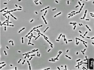
Ruminants have a four-chambered gut, and these microorganisms live primarily in the rumen. One particularly important bacterial genus that takes part in the degradation of cellulose is gram positive Ruminococcus (Figure 1). Ruminococcus bacteria break down the plant fiber into the monosaccharide glucose, which can then be further broken down through glycolysis. This symbiotic relationship enables ruminants to digest this fiber without having to encode for more enzymes in their own genomes to do this job. The relationship with microbes provides ruminants with about 15% of their caloric intake. The Ruminococcus genus, which includes Ruminococcus albus and Ruminococcus flavifaciens, is just one of many microbes living in the rumen. Others include Megasphaera, Fibrobacter, Streptococcus, Escherichia, Chytridiomycetes fungi, and methanogens. It is predicted that 70% of microbes in the rumen have yet to be identified.1 The proportions of microbes present vary greatly depending upon the diet of the ruminant. For this reason, the diets of the animals greatly impact their own health, and also effects consumers of their meat and the environment as a whole.
The Four Chambered Stomach and Function of Ruminant Microbes
The Stomach and Digestion
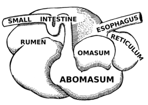
Ruminants have stomachs with four chambers; the rumen, the reticulum, the omasum, and the abomasum, as pictured in Figure 2. Food is first mixed with saliva and passed to the rumen, where it is mechanically broken down to smaller pieces and then passed to the reticulum. Here the food is further broken down and also separated from indigestible non-food items before it is formed into cuds. These cuds, or clumps of partially degraded food, are then regurgitated into the animal’s mouth, where it is re-chewed, and then re-swallowed back into the rumen. The majority of the anaerobic microbes that aid in cellulose breakdown inhabit the rumen, and during this step of digestion, fermentation begins. The partially digested food then moves to the omasum, where water, vitamins, and short chain fatty-acids from fermentation are absorbed into the animal’s body. pH is decreased and enzymes are released to further break down the material, before it is passed to the abomasum. The abomasum is the equivalent of the human stomach; material is further broken down and then passed to the small and large intestines where nutrients are absorbed before the waste is excreted.3 This process takes about 9-12 hours.3
Microbes in the Rumen
The rumen is an especially important chamber of the stomach, because it is here that cellulose is broken down into glucose, a sugar that can be used by the body. It is full of microbes that appear as soon as 38 hours after the animal’s birth.4 All three domains inhabit the rumen, but it is thought that bacteria are the most abundant, with 1011 viable cells/ml.10 Without the microbes, the ruminants would get about 15% less nutrients from their food. The ruminal bacteria themselves are also digested by the animals in their small intestines, and further serve as a source of nutrients.3 There are hundreds of different kinds of bacteria in the rumen which aid in this process, 85-95% which have yet to be cultured and 70% which have not yet been identified.1 Ruminococcus albus, and Ruminococcus flavefaciens are the principle cellulose degrading organisms.3 Despite their important role, cellulolytic bacteria are thought to only comprise 0.3% of the total ruminal bacteria population. 10
Diversity within the Rumen
Within the different areas of the rumen the types of microbes differ. Using PCR to amplify 16s rDNA sequences of microbes in the rumen, the fluid, solid, and epithelium (cellular layer lining of rumen) were determined to host different populations (Figure 3).1 The fluid had high levels of Cytophaga-Flexibacter-Bacteroides (67.5%), low G+C gram-positive bacteria (30%), and 2.5% proteobacteria. The solid was found to have the highest levels of low G+C gram positive bacteria (75.7%), 10.8% CFB, 5.4% proteobacteria and high G+C gram positive bacteria, and 2.7% spirochaetes. The epithelium had 94% CFB and 5.6% low G+C gram positive bacteria.1 This suggests that these microbes play specific roles in rumen function. The most abundant organisms can effectively degrade and colonize the plant material, and can utilize a wide range of substrates, thus outcompeting the other microbes.
Within the rumen, it is necessary for the microbes to be able to attach to the plant material. Fungi disrupt the exterior recalcitrant tissue of the feed creating holes to make it more susceptible to enzymes.9 Primary colonizing microbes then create glycocalyx-enclosed microcolonies, which are joined by secondary microbes from the ruminal fluid, forming the multispecies biofilm.4 This biofilm produces enzymes needed for the breakdown of cellulose. Using a complex metabolic pathway, these microbes turn cellulose into materials that can be used as energy sources by the ruminant animal as well as the microbe community.
Metabolic Pathway
Cellulose Structure
Cellulose plays an important structural role in plants but despite its natural abundance, it can not be broken down by many mammals. Cellulose is a homopolymer of glucose joined by β-1,4 linkages (Figure 4). Cellulose molecules associate with one another through hydrogen bonding and Van der waals forces to form microfibrils. These group together in parallel to form fibers, which are crystalline formations. This material makes up about 40% of plant cell walls.3 Ruminococcus, in the presence of branched-chain fatty acids (from the degradation of amino acids by different microorganisms) convert cellulose to glucose. This is particularly important because it is rare for organisms to be able to break down β-1,4 linkages. For this reason, certain cellulolytic species are predominant in herbivores so that they can optimize the nutrition obtained from cellulosic feed.5 Glucose is then further converted to acetate, pyruvate, and lactate through glycolysis and fermentation. In the process of this catabolic reaction, NAD+ is oxidized to NADH, producing H2 which methanogens in the rumen use with CO2, releasing CH4 into the environment.7
Attachment and Enzymatic Activity
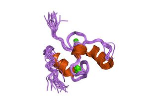
The degradation of cellulose occurs when the β-1,4 linkages are hydrolyzed by cellulase enzymes in Ruminococcus. A type of cellulase, endoglycosidase cleaves the disaccharide cellobiose from cellulose, and another type of enzyme, β-glucosidase hydrolyzes cellobiose and cellodextrins, producing glucose.3 Degradation is made possible by large, multienzyme complexes known as cellulosomes, pictured in Figure 5.5 The attachment of the complex to the cellulose substrate is coordinated in part by noncellulolytic microbes.9 Cellulose-binding proteins within the cellulase enzyme complex are thought to direct the attachment of the complex onto the crystalline cellulose structure.4 Scaffoldin or cellulosome-integrating proteins, large glycosylated proteins, can facilitate enzyme attachment, cellulose binding, or anchoring to the bacterial cell surface.5 These proteins have separate sections for binding and cleavage of polysaccharide bonds.5 The discovery of the cellulosome explains how anaerobic cellulolytic bacteria adhere so tightly to plant material, and why their enzymatic cellulase activity occurs primarily on the surface5. Alongside the cellulosome-structure, it is thought that Ruminoccus also uses a cellulose-binding protein type C (Pil) –protein mechanism that involves fimbrial structures that interact with cellulose. The importance of attachment can be seen in the fact then when Ruminococcus loses its ability to adhere to cellulose, it also loses its enzymatic ability.5
Factors that Effect the Microbe Population
The Effect of Feed Type
The population of microbes in the rumen is highly variable. The formation of biofilms is dependent upon proper levels of O2, temperature, Na+ levels, and rumen pH.4 Type of feed changes these factors and changes the microbe population. A poor forage diet results in a high level of fungi such as Chytridiomycete mycelia on food particles and motile zoo spores in the ruminal fluid. A high cellulose diet on the other hand leads to high levels of the cellulolytic bacteria, Ruminococcus.7 The type of forage can also effect the population. In an experiment, sheep were fed different diets with a forage concentrate ratio of 70:30 forage:concentrate, with either alfalfa hay or grass as forage. Samples were taken from the rumen 0, 4, and 8 hours after feeding. The relative abundances of microbes were most affected after 4 hours.6 The Ruminococcus concentration was not effected, but the hay diet promoted greater relative abundance of F. succinogenes and fungi compared to a grass diet.6 Following a dietary change from corn-silage to grass-legume hay, sequence variation in the dockerin module of the cellulosome occurred, but the N-terminus sequence, the location of the cohesion module, was conserved.10 Overtime, the microbes adapt to the type of feed that the animal normally eats, and the population becomes stable. Mature biofilms form that most effectively degrade the feed. Suddenly changing the feed can decrease their rate and extent of digestion, because it changes the biofilm’s ability to attach to the substrate, which is crucial for digestion.4
Feed Caveats
Small amounts of branched-chain fatty acids are also needed for cellulolytic bacteria to function, and these are produced by amino acid fermeters such as Peptostreptoccus anaerobius, which produce ammonia as a byproduct. For this reason it is important to limit protein intake, because too much ammonia can poison the animal.7 High levels of starch, which is found in most modern grains, can also be dangerous. The starch digestion leads to higher levels of fermenters that generate high levels of acidity and gases. This can cause starch bloat in animals, but can also lead to acid-resistant pathogens in the rumen, such as E. coli.7 In a unique example of rumen diversity, certain ruminants in Australia are poisoned by normally edible leguminous plants, due to a rare microbe in their gut that converts the amino acid mimosine to the toxic 4-hyrdoxy-4(H)-pyridone (DHP), a toxic goitrogen.8
Environmental Impact
The feed type given to ruminants is important for environmental reasons. High levels of H2 and CO2 from methanogens can lead to high global methane emissions. Release of acetate, butyrate, and propionate which are products of glycolysis can also be further broken down by soil microbes into CO and CH4 after excretion.3 This is hard to avoid since the number of electron donors (the reduced food molecules), far exceeds the available electron acceptors, so the excess hydrogen and carbon dioxide is utilized by the methanogens.7 Nevertheless, it is a big environmental issue since methane emissions is one of the leading causes of global warming.
Food Born Pathogens
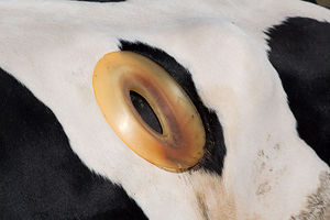
The prevalence of food born pathogens such as E. coli is an issue that can largely be controlled. In an experiment using a fistulated cow, a technique shown in Figure 6, it was determined that cattle fed more grain rather than hay have a lower gut pH and 106 fold higher levels of E. coli (Figure 7).2 This E. coli is acid resistant, which is dangerous because the high acidity of human stomachs that would normally kill the E. coli microbes once the animal meat was ingested is no longer effective. This has led to many more outbreaks of food born illness from ingesting meat as well as fruits and vegetables fertilized by manure. However, it was also found that once a grain-fed cattle is fed hay, the number of acid-resistant E. coli decreases very quickly. So, although it is unlikely that most commercially produced livestock will ever be fed hay diets, feeding them hay immediately before slaughter can reduce the risk of spreading E. coli.2
Conclusion
The degradation of cellulose in the stomachs of ruminants, made possible by microbes such as Ruminococcus, is crucial for the well-being and nutrition of the animals. The microbial population in the rumen is highly effected by the type of the feed the ruminant is given, so this is an important factor to consider in livestock production. Variance in the microbe populations can have detrimental environmental and health implications. Further investigation and identification of rumen microbes will be important in learning more about how the system works. More studies of how feed type, as well as possible other factors, effects the microbes will also be important to improve livestock production, prevent of food-borne pathogens, and decrease environmental impact
References
7Slonczewski JL and Foster JW. 2009. Microbiology: An Evolving Science. 2nd Edition. W.W. Norton, New York: 822-825.
Edited by KatiePruett, a student of Nora Sullivan in BIOL187S (Microbial Life) in The Keck Science Department of the Claremont Colleges Spring 2013.


