Dental Caries Prevention by Camellia sinensis
Mary Barker
Tooth Decay
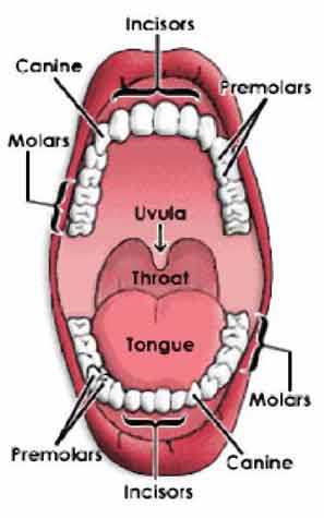
By the time the human mouth reaches maturity, most adult humans have thirty two teeth as is seen in Fig. 1. At the front of the mouth are incisors, the sharpest teeth, that are used to bite food and direct the food into the mouth. The canine teeth are located behind the incisors on either side of the mouth. They have long roots and grasp incoming food.
The pre molars are located even father back into the mouth. These teeth are wide and flat, equipped for grinding food before it is fully digested. Finally, the molars grind food into particles small enough to be swallowed and broken down in human’s digestive tracts.
It is very important to keep all of these teeth clean, and cavities are a real concern for humans in today’s society. After food enters the mouth, particles and bacteria are able to cling to the surface of the teeth. Many times, if the teeth are not brushed soon enough after eating, the bacteria can grow and form a film on the teeth.
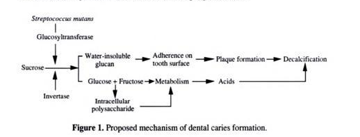
It is important to understand that cavities are not formed by the sugars consumed by humans, but instead by the bacteria that grow in the mouth if food particles remain on and between teeth for an extended period of time. Certain bacteria thrive in the conditions found in the human mouth. The warm environment and constant source of food both make the teeth and gums ideal locations for bacterial cultures to grow. Streptococcus mutans and Streptococcus sobrinus are two such bacteria commonly found in dental cavities. When they grow on the surfaces of teeth, they eat at the food particles and release acid as a waste product [5]. The path that this process typically takes is seen in figure 2.
This acid eventually builds-up, and breaks down the minerals in the teeth. Over time, when large amounts of acid have been released surrounding teeth, cavities begin to form. The acid initially leaves the surface of the tooth intact, while breaking down the enamel lying beneath the surface. When the tooth has enough damage, the surface also breaks down and a cavity is formed.
The most common means to prevent tooth decay are to consistently brush and floss the teeth. Flouride is usually present in toothpaste as a means to breakdown bacteria and prevent acid build-up. Flouride is also present in green and black tea, one reason why drinking tea can prevent tooth decay. In some areas, fluoride is also present in the public drinking water, although this is a topic of much debate recently [1].
Many Americans support the use of fluoride in their drinking water. This provides a way for humans to protect their teeth without making much of a personal effort, beyond drinking the water provided them from their faucets. It saves Americans millions in dental care costs every year [1]. In addition, those who support fluoridation argue that it helps all members of the community, especially the disadvantaged who are the most susceptible to dental problems. Supporters also point out that this is a long-term, effective, and health conscious use of tax dollars [1]. Some, however, disagree. Most critics of this process either are concerned about the possible health risks of consuming fluoride, or they are opposed to the fact that this decision has been taken out of their hands and as Americans they have no control over what they are drinking. These people argue that there have been studies showing fluoride causes considerable harm in biological systems, and can even be a factor in diseases such as cancer, infertility, and brain damage [1]. They also point out that much of Europe has removed the fluoride from their water systems. Finally, other critics are less concerned about the health effects of fluoride, and more disturbed by the lack of control they have over the chemicals in the drinking water they consuming. Should citizens have to buy water in order to ensure it is chemical free?
Bacterium Causing Tooth Decay
Ancient Japanese folklore tells how drinking tea leads to long life and clean teeth. At least the second part of this fable seems to be true. Recent research indicates that tea is able to counter some of the microorganisms, Streptococcus mutans, Streptococcus sobrinus, and Lactobacillus that can form plaque and biofilms on teeth, resulting in tooth decay.
Microorganisms from the genus Streptococcus are gram-positive bacteria. They have a round shape and frequently grow in chains. They are anaerobes that thrive in a complex culture. Streptococcus can cause many diseases, ranging from strep throat, to necrotizing fasciitis (flesh-eating disease) to the more mild tooth biofilm build-up [2]. Although not the most dangerous infliction caused by this genus, here the focus is on the latter condition and how to prevent it.
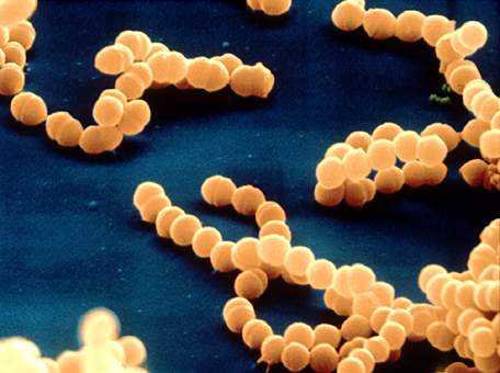
Streptococcus mutans (Fig. 3) is a species of Streptococcus that usually resides in the human mouth. It was discovered in 1924 by J K Clark and has recently been a topic of study due to several hundred unique genes that the bacteria has. These genes could potentially be a targeted in an attempt to kill the bacteria that cause tooth decay. S mutans is able to cling to the surface of teeth and feed on food particles, especially carbohydrates, that become trapped on and between teeth. The acid released by this bacteria is the leading cause of tooth decay in the world.
Streptococcus sobrinus is closely related to S. mutans, although it is less common in human beings. The two bacterial species are very similar and can only be distinquished with advanced technology and testing. In one study, the varying species were obtained through plaque samples, its DNA was amplified, and species were detected by PCR in a collection of school aged children. S. mutans was found 72.8% of the children tested, while S. sobrinus was only found in 61.1% of the subjects. [3]
Although S. mutans may be more common in the mouth, it has been shown that S. sobrinus has a greater impact on tooth decay. Students having both species had a significantly higher level of tooth decay than those with only S. mutans. The children with the most decay had both S. mutans and S. sobrinus present on their teeth. [3]
Lactobacillus are also a gram positive bacteria. They are organotrophs that develop in a rod-like shape. They can sometimes grow in clusters, and are especially prominent in tooth biofilm, where they thrive in the high sugar conditions. Lactobacillus’ main source of energy is found from metabolizing sugars into lactic acid, a process that makes them prime candidates for mouth dwellers. [4]
Tea
Tea is a drink derived from boiling the cured leaves of the Camillia sinensis plant. Camillia sinensis is grown in many places around the world, especially India and China. There are fours major types of tea than fall under the speciea C. sinensis; white, green, black, and oolong. In the study of dental caries prevention, green, black, and oolong are involved. Tea is consumed by billions of people around the world and after water is the most highly consumed beverage. [7]
Tea’s antioxidant properties are derived from the catechins compounds found in tea leaves. Catechins are flavoniods found mostly in the leaves of tea, although they are also in wine, chocolate and fruits.
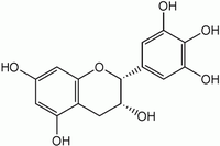
Tea’s antioxidant powers have a plethora of healthy effects, including tumor suppression and cancer prevention. Epicatchin gallate (ECG), seen in figure 4, is a catechin found in green tea. It can prevent the formation of carcinogens in animal studies [6].
Other flavonoids also derived from tea also have varied health benefits. They protect the heart against disease through their ability to prevent the oxidation of some lipoproteins in macrophages [6]. Saponins extracted from green tea have an anti-inflammatory effect, studied in rats because the saponins decrease the ability of glucose and salt to be absorbed in the intestine.
Tea has been studied in experiments involving liver damage and HIV and are commonly used in household products such as mouthwash and deodorant [6].
Effects of Tea on Dental Caries
Looking at the molecular processes involved in teeth decay can help explain the nature of dental caries prevention. Dental caries is a gradual process that begins with the accumulation of bacteria in the mouth, usually caused by prolonged exposure of the teeth and mouth to sucrose. This process has three basic steps.
1. Bacteria, usually from a food source, attached to the teeth.
2. Glycocalyx is formed when glucosyl transferase, a bacterial enzyme, reacts with sucrose
3. Formation of biofilm, as bacteria metabolize carbohydrates and produce acid that eventually decays the tooth.[5]
Green, black, and oolong teas have specifically been studied in relation to teeth health. Green and black tea share many similarities, yet differ slightly in the structure of their catechins. Green tea has simple catechins with a lower molecular weight than black catechins. Many black catechins have been oxidized through fermentation and have a higher molecular weight, although some remain simple. [5] Catechins can remain in the mouth for up to sixty minutes after tea has been consumed.
A cup of green tea contains three times as many catechins than a brewed cup of black tea. [5]. Oolong tea is an intermediate between these two types, containing both simple and fermented catechins.
There have been many studies exploring the molecular methods behind the ability of tea to fight tooth decay. It has been discovered that these teas actually attack each of the three basic steps to dental caries. The first step is the adhesion of the bacteria to the tooth. Green and black tea extracts inhibits the ability of S. mutans to bind to the tooth surface. In addition, Oolong extracts have been found to have an affinity for proteins. When the oolong binds to the surface proteins of the tooth, they prevent other bacteria from attaching there as well. [5]
The second step of tooth decay involves the production of glycocalyx, a sugar coat produced from oral bacteria. Green and black tea contains catechins that reduce the level of enzyme activity in glucosyl transferase. This lowers glycocalyx levels on the tooth.
Green, and especially black tea have been found to prevent salivary amylase in S. mutans. The catechins in black tea, epigallocatechin gallate (EGCG) having a heavier molecular weight are especially detrimental to amylase activity. This is caused by the ability of EGCG to breakdown the membranes of the oral bacteria.
Once a biofilm has been established on the surface of teeth, it becomes very hard to eradicate. Biofilms are strong evolutionary communities that join bacteria together to provide structure and support, as well as other benefits including exchange of antibiotic resistance. It is much easier and more effective to break down growing bacteria before they reach this stage.
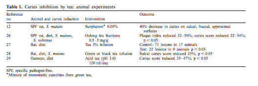
There is not a substantial amount of evidence concerning the specific reasons that tea is able to harm the bacteria in the human mouth. It has been found, however, that in trials in which tea was incorporated in the diets of certain animals, rats and hamsters specifically, there was a reduction in the amount caries on the teeth surfaces, as seen in the above table. The effects of tea on rat caries is seen in figure 5. Rats responded especially well to the tea extract treatments. In each treatment the caries score decreased significantly. [2]
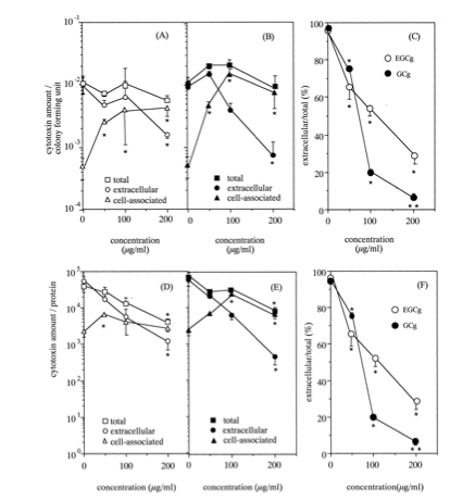
Although there is not a lot of information about the mechanism through which tea extracts prevent tooth decay, there is some knowledge on the effects of tea on other bacteria. In a 1999 study by Sugita-Konishi et al. the means through which green tea is able to inhibit toxin release in E. coli was studied. This study focused on Escherichia coli, which is a commonly known food contaminant. It is found in unwashed vegetables and meats and through digestion moves into the human digestive tract, especially into the large intestine. Here, the bacteria are able to colonize and release vero toxins (VTs) which cause gastroenteritis [8]. In this study, samples of the bacteria were place on a soy broth and six tea catechins were isolated from green tea. The six catechins were each placed in a separate culture along with the bacteria, and samples from each group were taken at set times over a twenty four hour period. The amount of vero toxins in each sample was determined using reversed passive latex agglutination [8]. All six of the catechins did not have significant inhibition effects on the ability of the E. coli to produce the toxins, however two did. ECGg and GCg completely inhibited the production of vero toxin 1 and, at higher concentrations, vero toxin 2 as well. As can be seen in figure 6 these two catechins significantly decreased the percentage of extracellular toxins, when compared to a control group lacking catechins. So, what is it about these two specific catechins that prevents the activity of bacteria?
The answer lies in the structure if EGCg and GCg. Both of these catechins, and none of the others studied, had at the 3-OH position and the hydroxyl group at the 5P-position the galloyl moiety [8]. It is believed that this is necessary to prevent vero toxin excretion in E. coli cells. The researchers then preformed father study to determine if these two catechins were also able to supress production of another periplasm protein, maltose binding protein (MBP).
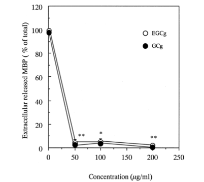
MBP is a part of the maltose system of E coli bacteria, and is responsible for the uptake and catabolism of maltodextrins from the extracellular environment. ECGg and GCg were both shown to significantly inhibit the release of this protein as well, as can be seen in Figure 7. Here, at catechin concentrations above fifty micrograms per milliliter the release of MBP is almost entirely supressed . This indicates that something in these catechins prevents the general release of a wide-range of periplasmic proteins, not only vero toxins [8]. The catechins most likely interfere with the ability of these proteins to exit the extracellular membrane of E. coli cells.
In much the same way, tea catechins are probably able to prevent the release of toxins from in dental caries bacteria. The catechins are able to prevent the release of Glycocalyx from the teeth binding bacteria. This would prevent them from being able to bind to the teeth surface and form biofilms. The catechins with a galloyl moiety would prevent the excretion of acids from the bacteria as well, preventing tooth decay. Overall, the ability of catechins to prevent toxic release in E. coli cells provides a basis for us to assume a similar mechanism in the prevention of dental caries by tea extracts.
Conclusion
In today's world, humans are bombarded daily with high sugar food and drinks. From soda pop to brownies, humans consume more sugar than our bodies want or need. Just one side effect of such consumption is unhealthy teeth. With our teeth being such a prominent aspect of our physical appearance, it is no surprise that oral cleanliness is important.
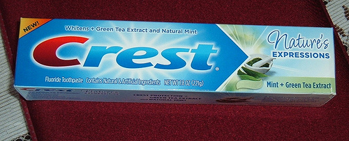
Humans have many ways of keeping teeth looking bright and healthy. Brushing and flossing is common, along with more extreme methods of teeth whitening and bleaching. However, drinking tea is another method that could be employed by humans to prevent teeth decay. Studies have shown that countries in which tea drinking is widespread, such as India, Japan, and China, there is a lower incidence of dental problems. [3] Many major toothpaste companies have taken the positive effects of green tea into consideration, and products such as green tea infused toothpaste are now on the market, for example the Crest toothpaste seen in figure 8.
Additional research is needed to determine the exact methods employed by green and black tea extracts to break down and inhibit the activity of S. mutans, S. sobrinus, etc. One exciting future research prospect is the ability of S. mutans to produce certain genes specific to this species. [2] If these genes could be identified and examined more closely, it is possible that antibodies could be created to target this specifically. This could lead to treatments and prevention of bacterial oral disease that are more effective and less time consuming than brushing and flossing.
References
4. Faro, James. "Lactobacillus acidophilus." 1999
Edited by student of Joan Slonczewski for BIOL 238 Microbiology, 2009, Kenyon College.
