Digestion in oysters
Introduction
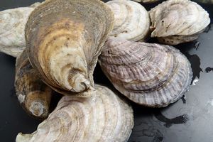
The Eastern oyster (Crassotrea virginica) is a marine bivalve mollusk.[1] Eastern oysters populate the Atlantic coastal waters of North America and the West Indies.[1] Oysters form beds by rereleasing spawn that settle and mature on or near existing oysters.[1] Oyster beds serve dually as social structures and physical structures for habitation.[1] Adult oysters are sessile, meaning they do not move from their anchorage, and they feed from their positions in the beds.[2] Like many other bivalves, oysters are filter feeders, and they subsist on phytoplankton.[1] Eastern oyster reproduction is synchronized to occur in early spring in anticipation of algal blooms that follow later in the season.[1] Oysters have developed anatomical structures and physiological processes that are highly specialized to filter particles from the water surrounding them.

Structures involved in feeding
Labial palps: The Palps are the organs that flank the mouth of the oyster.[3] They have a smooth outer level, muscular-connective tissue middle layer, and a ridged and ciliated inner layer, which faces the gills.[3] The grooves and cilia of the labial palps are responsible for removing excess material and detritus from the food tracts of the gills.[3] In the Eastern oyster, the gills also participate in particle rejection.[3] The cilia are organized by three functional tracts: rejection tracts, oral acceptance tracts, and resorting tracts.[3] If particles are directed into the oral acceptance tracts they are passed to the mouth.[3] If they are directed into the rejection tracts they are expelled from the oyster as pseudofeces, i.e. food waste created prior to actual ingestion.[3] Lastly, if particles are directed to the resorting tracts they are recirculated through the process, and in this the resorting tracts serve as a means of volume control for the other tracts.[3]
Gills: The gills of the Eastern oyster are of a plicate, heterorhabdic pseudolamellibranch construction.[3] This means that the gills are lined with cilia that are capable of both selecting and rejecting particles for absorption.[4] This selection process works in tandem with the palps and is based on particle size.[4] Cilia in the gill filaments move water through the gills.[3] As the water passes through the entrance of the interfilament spaces it is filtered by lateral cilia that contain mucocytes.[3] These mucus-coated cilia trap particles from the water that are then transported either to a mucus string near the palps or through plical troughs into the dorsal ciliated grooves.[3]
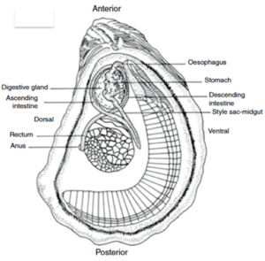
Esophagus and Stomach: Particles which enter the digestive tract pass through the esophagus and into the stomach.[3] The esophagus does not serve a digestive role and is merely involved in transportation.[3] The stomach secretes a crystalline style, or gel, comprised of digestive proteins and structural sugars.[3] Chitinase is found in high concentrations in the stomach because chitin is a component of cell walls in algae, a major food source of oysters.[3] The style is secreted by the style sac, but is not itself a permanent structure.[3] The stomach wall also secretes digestive enzymes, even when the organism is not feeding.[3]
Digestive Glands: Food is also processed intracellularly in the digestive glands, which are connected to the stomach and intestines.[3] The digestive glands absorb food through small tubules that contain digestive cells, basophil secretory cells.[3] The digestive cells digest the food material intracellularly and send nutrients into the hemolymph system and store the waste.[3] When they reach capacity they burst and the waste travel to the intestine. The basophil secretory cells appear to be involved in enzyme production.[3]
Intestine: The intestine is primarily involved with temporarily storing and eliminating waste, however some digestion does occur in the organ.[3] Extracellular digestion occurs here, as does intracellular digestion by wandering hemolymph cells.[3] The organ is also capable of nutrient transport across the membrane of the lumen.[3]

Genetics
Oysters vary widely by species, and even within a species they can produce an array of different phenotypes from genetic variance. This is due in large part to their lack of migration, as once a bed is separated – by geography, environmental conditions, or other factors – it becomes a naturally isolated population. How genetic change has led to evolution in the digestive system remains a topic for further research. However the distinct complex morphology of the digestive system offers a phylogenic history already. For instance, scallops have been found to have shared adaptations for particle selection in oysters that are lacking in the more deeply branching mussels.[4][3]
Microbiome
Bacteria associated with the digestive style were identified as early as 1882, however their function remains a mystery.[3] Recent recognition of the roles that microbiomes serve in the digestive systems of other animals has led to renewed interest in the microbial aspect of the digestive style and other digestive organs. Several recent studies have sought to observe the microbiomes of Eastern oysters and to explore what role the microbiome plays in health and metabolism within the oyster. Studies have shown Eastern oysters to have microbiomes with bacterial compositions distinct from the surrounding soil environment. In fact, one study found distinct microbiomes exist between the stomach and intestinal organs within the same organisms.[5] The same study also found significant difference between phylogenetic compositions of the microbiomes of two geographically separated Eastern oyster populations, one sampled from Hackberry Bay, LA and the other from Lake Caillou, LA.[5] It is not understood at this time whether the difference in phylogenetic composition of oyster microbiomes contributes to a difference in symbiotic function of the microbiome, or whether the microbiome is necessary for any regular metabolic function within oysters.[5] Further research is needed to determine what role, if any, oyster microbiomes play in digestion.
Conclusion
The anatomy and physiology of digestion in oysters is well understood, however research is lacking on what role bacteria may play in digestion. Furthermore while the complex morphologies of oyster digestive organs have offered insight into their evolution and phylogeny, a better genetic understanding is required to more accurately reflect this.
References
- ↑ 1.0 1.1 1.2 1.3 1.4 1.5 https://animaldiversity.org/site/accounts/information/Crassostrea_virginica.html Osborne,Paula. Crassostrea virginica. University of Michigan, Ann Arbor. 2014
- ↑ https://www.fisheries.noaa.gov/species/eastern-oyster
- ↑ 3.00 3.01 3.02 3.03 3.04 3.05 3.06 3.07 3.08 3.09 3.10 3.11 3.12 3.13 3.14 3.15 3.16 3.17 3.18 3.19 3.20 3.21 3.22 3.23 3.24 3.25 3.26 Gosling, Elizabeth. Marine Bivalve Molluscs. Second edition, John Wiley & Sons LTD. 2015.
- ↑ 4.0 4.1 4.2 https://peter-beninger.com/Localization%20of%20qualitative%20particle%20selection%20sites.2004.pdf Beninger, Peter, Priscilla Decottignies, and Yves Rincé. Localization of qualitative particle selection sites in the heterorhabdic filibranch Pecten maximus(Bivalvia: Pectinidae). Marine Ecology Progress Series, MARINE ECOLOGY PROGRESS SERIES, Mar Ecol Prog Ser Vol. 275: 163 –173, 2004.
- ↑ 5.0 5.1 5.2 https://journals.plos.org/plosone/article?id=10.1371/journal.pone.0051475 King, Gary, Craig Judd, Cheryl R. Kuske, Conor Smith. Analysis of Stomach and Gut Microbiomes of the Eastern Oyster (Crassostrea virginica) from Coastal Louisiana, USA. Plos One, 2012.
Edited by [David Hamilton], student of Joan Slonczewski for BIOL 116 Information in Living Systems, 2019, Kenyon College.
