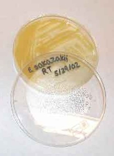
Enterobacter sakazakii
A Microbial Biorealm page on the genus Enterobacter sakazakii
Classification
Higher order taxa
Bacteria; Proteobacteria; Gammaproteobacteria; Enterobacterials; Enteribacteriaceae
Species
Enterobacter skazakii
Description and significance
Enerobacter sakazakii is described as ubiquitous and opportunistic pathogen that currently contaminates a wide spectrum of foods and poses a lethal threat to neonates, the elderly and persons with immune deficiencies. This species is known to be gram negative, produce a yellow culture, have a rod shaped body structure that aproximatly measures 3 by 1 micrometers which is highly flagellated (peritrichous) and can produce a protective biofilm. This biofilm has allowed this species to establish it self as food pathogen in most food industries and handling facilities. E. sakazakii is comprised of 134 strains which been recorded to cause death in neonates since the 1920’s and has since caused sporadic deaths for the last 50 years. It was officially distinguished as a species in the late 1970’s were it was isolated and found to cause life threatening neonateal meningitis, septicemia and necrotizing enterocolitis. Currently, there is still a great deal that is not known about E. sakazakii such as its natural habitat, genomic sequence and virulence factors. (1)(2)(4)(5)(7)
Genome structure
The genomic content of Enterobacter sakazakii is currently being sequenced at Washington University (WashU) and is being funded by the National Institute of Allergy and Infectious Diseases (NIAID) and Nation Institutes of Health (NIH). Sequencing methods consist of plasmid and fosmid libraries which has an expected error rate of less than 1 per 10 kb. The particular strain being sequenced was isolated from a hospital’s infant formula stock that was previously used to nourish neonates. (8)
As of 2006, strains of E. sakazakii have been segregated by researchers for classification purposes via: amplified fragment length polymorphisms fingerprints (f-AFLP), ribopatterns, full length 16S r-RNA gene sequencing and DNA-DNA hybridizations with different strains of E. sakazakii to compare relatedness between the species. These studies have lead current researchers to propose a new genus Cronobacter, but are reluctant to reclassify the bacteria as of yet, due to the concern that it might jeopardize health protection measures that were placed in 2001 by the FDA. The proposed reclassification of E. sakazakii designates four new species along with one genomospecies and two subspecies in the family Enertobacteriaceae, they are as follows: Cronobacter sakazakii subsp. sakazakii, Cronobacter sakazakii subsp. malonaticus, Cronobacter muytjensii, Cronobacter dublinensis, Cronobacter turicensis, and the genomospecies Cronobacter. Currently, the National Center for Biotechnology Information (NCBI) lists both Cronobacter and Enterobacter as the taxonomic genus but lists the comment that the former is an “unpublished name…was not validly published at time of submission of corresponding sequence…” (1)(9)
Cell structure and metabolism
E. sakazakii is a Gram negative, rod shaped bacterium measuring approximately between 3 μm by 1μm in size and has been found to be peritrichous or highly flagellated, thus being motile. It has historically been describe since as early as 1929 when it was characterized by producing a yellow culture and causing septicemia in infant(s). Recent experiments have primarily focused on ways to prevent it from entering human food sources or treating foods once it has been contaminated. The heterogeneity between the E. sakazakii species have made these experiments difficult to analyze due to the 134 strains that often share metabolic pathways but, tend differ in survival strategies. Nevertheless, studies show some interesting similarities amongst the strains which portray a highly versatile and hardy microbe that produces capsule, biofilm and uses an adhesive behavior that is still unclear. The capsule and biofilm have been shown to reduce the efficacy of various common sanitizing methods such as: UV-light radiation, high osmotic pressures, heat, dry conditions, starvation, low pH, detergents, antibiotics, phagocytes, antibodies and some bacteriophages. (2)(4)(6)(7)
The adhesive behavior of E. sakazakii strains is just as impressive but until only recently has it had thorough research. Adherence has been recorded on inorganic and synthetic surfaces such as: plastics, silicon, PVC, polycarbonate, glass, stainless steel and in public water systems. Adhesion to living surfaces such as in a pathogenic capacity has been shown to differ between strains but all share the trait of being independent of fimbriae structures and require a metabolically active host cell for adhesion. (6)
Strains in this genus have been shown to have metabolic pathways that are characterized as being facultative anaerobic with oxidase negative and catalase positive capabilities. Substrates that can be metabolized for energy include nitrates in nitrate reduction reactions, sugars, amino acids and various lipophilic molecules.(1)
Ecology
Described as ubiquitous and opportunistic bacteria since its official discovery in 1980, research involving man-made food processing plants has been a major focus of research since it was first associated with milk-based powdered infant formulas. Research has since been used to include other food industries and it has established that around the globe E.skazakii can be cultured from a huge spectrum of food sources, which briefly include: vegetables, fruits, legumes, grains, a variety of meat containing products, milk and drinking water both from tap and bottled. These food sources listed include frozen, raw, cooked and pasteurized foods that have been found at retail levels throughout the world. (2) The natural environment of E. skazakii and its role is still unknown.
Pathology
E. sakazakii is a food borne pathogen can produce severe illnesses and death to persons with immunological deficiencies such as neonates, the elderly and persons with severe underlying diseases. In these populations E.sakazakii can successfully colonize, establish and ultimately produce disease. Virulence factors are still largely unknown and pathogenicity mechanisms have only begun to be researched. The adherence aspect of E.sakazakii virulence, maybe furthest along with regard to determining its properties pathenogenicity. (2)(6)
In humans, E. sakazakii has been found to affect specifically the vascular system the gastrointestinal system and the nervous system. Establishment in the human vascular system causes bactereaemia and/or sepsis which often proceeded by colonization beyond the blood brain barrier which results in ceribro spinal fluid infection and meningitis; this progresses into intracerebral infarctions, brain abscess and/or cyst formation resulting in central nervous system deterioration. Necrotizing enterocolitis (NEC), also associated with E. sakazakii infection, and is currently the most common gastrointestinal emergency in neonates, it characterized by necrosis of the gastrointestinal lumen.
Once infected, neonates have been found to have a mortality rate of 40 to 80% when infected and a 20% chance that survival is accompanied with serious neurological complications such as hydrocephalus, quadriplegia, brain abscess and retarded neural development (3)(5)(6).
Application to Biotechnology
Determining the complete genome sequence and allowing time for bioinformatic analysis is necessary in order to identify any practical uses for this species. (5)
Current Research
One of the more crucial types of research concerning E. sakazakii has been in developing novel ways in identifying it in food sources and in the general environment. Fatty Acid profiling seems to hold great promise as rapid GC-FID method that can identify and differentiate food pathogens by up to the species level in clinical, environmental, and food samples. This method of identification analyzes fatty acid compositions of whole bacteria cells to determine it’s (saturated: monounsaturated: cyclopropane) fatty acid ratios. A data base with the ability to identify all 134 strains of E. sakazakii fatty acid profiles has been developed by researchers and can produced reliable species level identification in about 24hrs.(7)
Novel ways of treating contaminated foods has also been an area of current research. Typical commercial sterilizing techniques such as UV radiation and several chemical disinfectants have been known to have a reduced efficacy towards E. sakazakii but, recent experiments have shown great promise in the use of bacteriophages. Four new lytic bacteriophages were isolated from a sewer treatment plant in Switzerland and have shown great efficacy in inhibiting outgrowth in bacteria cultures isolated from infant formula after only 4 hours inoculation; two phages in particular have even exhibited host specificity towards E. sakazakii. Applications of Listera phages as a non-destructive biocontrol method has been approved for use in foods since 2006 by the FDA, as a replacement or supplement to conventional controls; thus, the proposition of using phages to control this bacteria’s ubiquity in the food industry holds promise. Further information pertaining to E.sakazakii such as the genomic sequence, bioinformatic data as well as further tests with the new phage and increasing the number of potential phages is required if this bio-control is ever going to have any practical applications. (4)(5)(7)
New hygienic practices have been an area of research that has progressed slower compared to other areas due E. sakazakii biofilms and adhesive properties. It has been know for quite some time that food contamination for these bacteria often originates from using conventionally clean utensils and equipment for the preparation of infant formula in industrialized production, hospitals, day-care centers, food service kitchens and at home. Commercial liquid chemical disinfectants that so far have shown varying digress of efficacy. This is further complicated because the resiliency of E. sakazakii to disinfectants varies from strain to strain. As of now, data has shown that the best way to prevent biofilms from proliferating on food utensils has been to remove all trace amounts of infant formula or food, then follow with a disinfectant. The theory is that infant formula acts as a protective nourishing layer when left on utensils; therefore, removing all food particles will increase lethality of disinfectants. (2)(4)
References
(8)Washington University in St. Louis – Genome Sequencing Center : Enterobacter sakazakii.
(9)NCBI: Enterobacter sakazakii.
Edited by student of Rachel Larsen and Kit Pogliano

