Euglyphids
A Microbial Biorealm page on the Euglyphids

Classification
Higher order taxa:
Eukaryota; Cercozoa
Species:
Euglypha strigosa, E. compressa, E. patella
|
NCBI: Taxonomy Genome |
Description and Significance
Euglyphida is a monophylitic genus, sharing a lineage with its sister group, sarcomonads. They belong to the group Cercozoa, which was defined by Thomas Cavalier-Smith in 1998. There are roughly 40 known species.
Genome Structure
At present time, there has not been extensive research done on the genome structure of Euglyphids.
Cell Structure and Metabolism

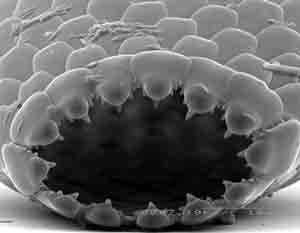
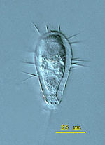
Euglyphida are unicellular organisms lacking flagella, cytosomes, and centrioles. They have silica scales; these are shaped like ovals. There are about 150 scales on each organism, making up a test (shell). These scales are formed by surface-excretion. Scales are held together with a sticky secretion called pseudochitin. This test is often transparent, and many are equipped with spines. The organism can retract into this case if it is disturbed or threatened. Euglyphida move and feed with filipodia.
Euglyphida are heterotrophic organisms. They mainly feed on bacteriam, as well as plant detritus.
Euglyphida are asexual organisms. During reproduction, shell plates are constructed in the Gogli bodies, as well as pernicular regions, of the parent cell. They pass along microtubule pathways during the binary fission process. They are positioned in the daughter cell by microtubule and microfilament systems.
Ecology

Euglyphida are mainly freshwater organisms. However, there are also marine species, as well as species that are terrestrial, living on mosses. Because they feed on plant detritus, Euglyphida have an important role in the decomposition process. They can also be found in waste water, feeding off bacteria. Euglyphida can also be found in moist soil, peat, or lake sediments.
Euglyphida can serve as bioindicators for aquatic environments; they can also be used to understand what specific geographic areas were like earlier in the formation of the planet.
References
Engitech Inc. Environmental Training. Activated Sludge Microorgaisms.
Micrographia. The Testate Amoebae.
Microscope: Information About Microbes.
