File:Nematopsis1.gif
From MicrobeWiki, the student-edited microbiology resource
Size of this preview: 506 × 599 pixels. Other resolution: 929 × 1,100 pixels.
Original file (929 × 1,100 pixels, file size: 876 KB, MIME type: image/gif)
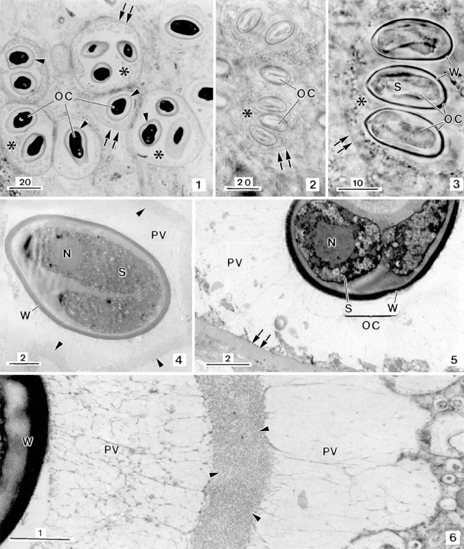
Light microscopy of the apicomplexan gregarine, Nematopsis gigas n. sp., a parasite found in the mantle tissue of the gastropod Nerita ascencionis (all scale bars in μm). Semithin section showing some host phagocytes (*) each containing some oocysts (OC) located in a lighter area (parasitophorous vacuole). Between each oocyst and the periphery of the parasitophorous vacuole a dense structure (arrowheads) was observed. The “phagocyte” walls are indicated by double arrows.
File history
Click on a date/time to view the file as it appeared at that time.
| Date/Time | Thumbnail | Dimensions | User | Comment | |
|---|---|---|---|---|---|
| current | 15:05, 12 December 2016 | | 929 × 1,100 (876 KB) | Suppleej (talk | contribs) | Light microscopy of the apicomplexan gregarine, Nematopsis gigas n. sp., a parasite found in the mantle tissue of the gastropod Nerita ascencionis (all scale bars in μm). Semithin section showing some host phagocytes (*) each containing some oocysts (... |
| 15:03, 12 December 2016 | Error creating thumbnail: File missing | 929 × 1,100 (876 KB) | Suppleej (talk | contribs) | Light microscopy of the apicomplexan gregarine, Nematopsis gigas n. sp., a parasite found in the mantle tissue of the gastropod Nerita ascencionis (all scale bars in μm). Semithin section showing some host phagocytes (*) each containing some oocysts (... |
You cannot overwrite this file.
File usage
The following page uses this file:
