File:Nematopsis2.gif
From MicrobeWiki, the student-edited microbiology resource
Size of this preview: 487 × 599 pixels. Other resolution: 797 × 981 pixels.
Original file (797 × 981 pixels, file size: 764 KB, MIME type: image/gif)
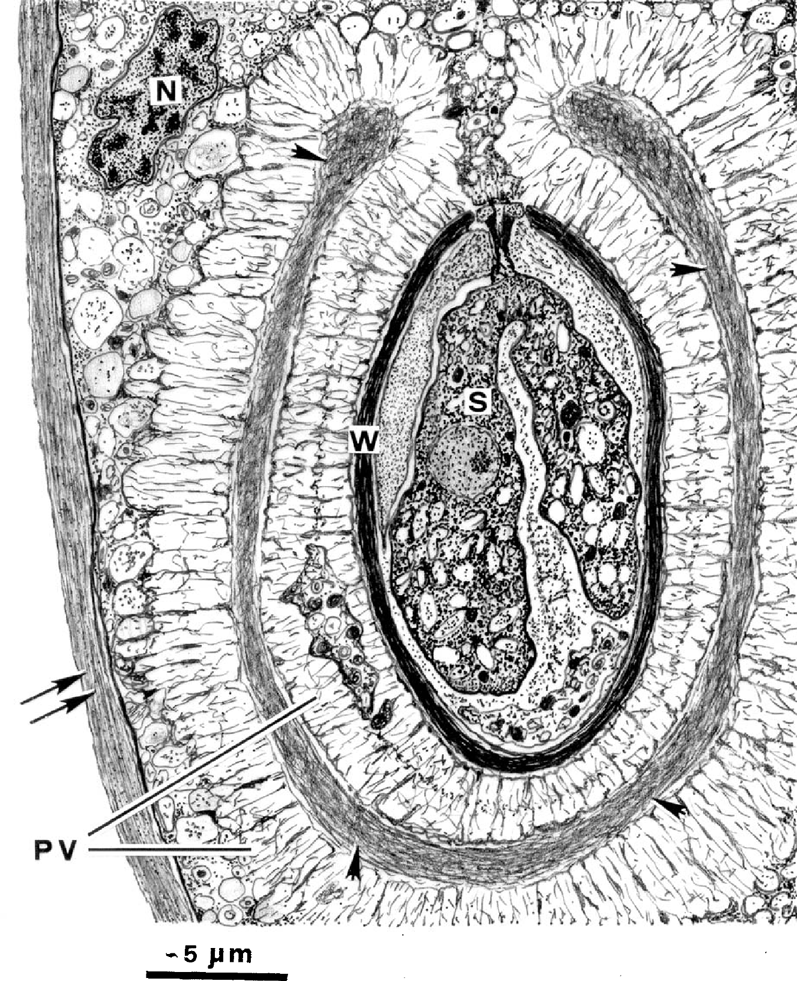
Schematic drawing of a longitudinal section of an oocyst of Nematopsis gigas n. sp. and the surrounding structures of the parasito-phorous vacuole (PV). Note the numerous anastomosing microfibrils of the PV, some of which form a dense and complex network (arrowheads). The phagocyte has an eccentric nucleus (N), a vesicular cytoplasm, and is surrounded by the “phagocyte” wall (double arrows). (2) Copyright of Journal of Eukaryotic Microbiology, reprinted with permission from the author.
File history
Click on a date/time to view the file as it appeared at that time.
| Date/Time | Thumbnail | Dimensions | User | Comment | |
|---|---|---|---|---|---|
| current | 15:06, 12 December 2016 | | 797 × 981 (764 KB) | Suppleej (talk | contribs) | Schematic drawing of a longitudinal section of an oocyst of Nematopsis gigas n. sp. and the surrounding structures of the parasito-phorous vacuole (PV). Note the numerous anastomosing microfibrils of the PV, some of which form a dense and complex netwo... |
You cannot overwrite this file.
File usage
The following page uses this file:
