Human immunodeficiency virus
A Viral Biorealm page on the genus Human immunodeficiency virus

Baltimore Classification
Higher order taxa
Viruses; Retro-transcribing viruses; Retroviridae; Orthoretrovirinae; Lentivirus; Primate lentivirus group; Human immunodeficiency virus (HIV)
Description and Significance
HIV is the causative agent of Acquired Immunodeficiency Syndrome (AIDS). AIDS is a severe, life-threatening disease that represents the late clinical stage of infection with the HIV. 2.5 million people died of AIDS in 2005 alone, and estimates place the number of people living with HIV/AIDS at 38.6 million. HIV/AIDS has claimed more than 25 million lives since 1981.
The Centers for Disease Control reported cases of Pneumocystis carinii pneumonia and Kaposi's sarcoma in otherwise healthy young male homosexuals in 1981. Until then, pneumocystis carinii was mainly known to occur in immunodepressed patients after organ transplants or suffering from congenital immunodeficiencies. Soon thereafter, the same condition was seen in IV drug abusers, haemophilliacs and babies of IV drug abusing mothers. These patients had profound immunosuppression due to the depletion of T4 helper lymphocytes and the name 'acquired immunodeficiency' was coined for this syndrome. Epidemiological studies have now established that the disease is infectious and can be transmitted by sexual intercourse, blood or blood products. The lymphocytes of patients died early, creating a difficulty in isolating the virus. Montagnier and Gallo eventually isolated the virus in 1984 and HIV-2 was isolated in 1986 from West Africa. HIV-1 and HIV-2 do not cross-react serologically with each other in screening tests. (sources: Avert, Virology-Online)
Genome Structure
The genome of HIV-1 is dimeric, unsegmented and contains a single molecule of linear. The genome is -RT and is positive-sense, single-stranded RNA. The complete genome is fully sequenced and of one monomer 9200 nucleotides long. The genome has terminally redundant sequences that have long terminal repeats (LTR) of about 600 nt. The 5'-end of the genome has a methylated nucleotide cap with a sequence of type 1 m7G5ppp6'GmpNp. The 3'-terminus has a poly (A) tract and has a tRNA-like structure and accepts lysin. Two copies of the genome are present in the virion in a dimeric configuration with two copies per particle being held together by hydrogen bonds to form a dimer. (source: ICTV db Descriptions)
Virion Structure of a Human immunodeficiency virus
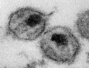
The virions of an HIV-1 consist of an envelope, a nucleocapsid, a nucleoid, and a matrix protein. The virus capsid is enveloped. The virions are spherical to pleomorphic and measure 80-100 nm in diameter. The surface projections are small, at 8 nm in length, but densely dispersed and there are inconspicuous spikes that cover the surface evenly. The nucleoid is concentric while the core is rod-shaped or truncated cone-shaped. (source: ICTV db Descriptions)
Reproduction Cycle of a Human immunodeficiency virus in a Host Cell
The replication of HIV can only take place inside human cells. The process typically begins when a virus particle bumps into a cell that carries a special protein called CD4 on its surface. The spikes on the surface of the virusparticle stick to the CD4 to allow the viral envelope to fuse with the cell membrane. HIV particle contents are then released into the cell, leaving the envelope behind.
The HIV enzyme reverse transcriptase converts the viral RNA into DNA, which is compatible to human genetic material, when the virus is inside the cell. This DNA is transported to the cell's nucleus, where it is spliced into human DNA by the HIV enzyme integrase. The HIV DNA is known as provirus after it is integrated.
HIV provirus may lie dormant within a cell for a long time but when the cell becomes activated, it treats HIV genes in much the same way as human genes. First, it converts them into mRNAs using human enzymes. The mRNA is then transported outside the nucleus and is used as a blueprint for producing new HIV proteins and enzymes.
There are complete copies of HIV genetic material among the strands of mRNAs produced by the cell. These gather together with newly made HIV proteins and enzymes to form new viral particles, which are then released from the cell. The enzyme protease plays a vitla role at this stage of HIV's life cycle by cutting down long strands of protein into smaller pieces, which are used to construct mature viral cores.
The newly matured HIV particles are ready to infect another cell and begin the replication process all over again. The virus quickly spreads through the human body in this fashion. (source: AVERT)
Viral Ecology & Pathology
The earliest unambiguously identified HIV-antibody positive serum stems from Kinhasa, Zaire dating back to 1959. HIV infection spread unrecognized in the 1960s and 1970s before it was finally recognized in 1981. The spread of the virus has been phenomenal thereafter, and close to 40 million people are estimated to be infected with the virus.
The profound immunosupression seen in AIDS is due to the depletion of T4 helper lymphocytes. HIV is present at a high level in the blood immediately after exposure. It then settles down to a certain low level set-point during the incubation period that lasts from 3-8 weeks. During the incubation perid, there is a massive turnover of CD4 cells as the CD4 cells killed by HIV are replaced rapidly and efficiently. The immune system eventually succumbs and AIDS is developed when killed CD4 cells can no longer be replaced, as witnessed by high HIV-RNA, HIV-Antigen and low CD4 counts.
Activated cells that become infected with HIV produce virus immediately and die within one or two days. The vast majority of viruses present in the plasma can be attributed to the short-lived, activated cells. It takes approximately 1.5 days to complete a single HIV life-cycle. Resting cells that become infected produce virus only after immune stimulation and these cells have a half-life of at least 5-6 months. Some cells are infected with defective virus that cannot complete the viral cycle. Such cells survive for a long period of time and have an estimated half-life of 3-6 months. (source: Virology-Online)
