Leukocytozoon
A Microbial Biorealm page on the genus Leukocytozoon

Classification
Higher order taxa
Eukaryota; Alveolata; Apicomplexa; Haemosporida
Species
Leucocytozoon smithi
Leucocytozoon simondi
Leucocytozoon caulleryi
|
NCBI: Taxonomy Genome |
Description and Significance
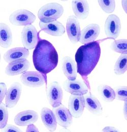
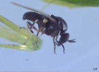
Leucocytozoons are parasites which cause leucocytozoonosis in birds. They are the most important blood protozoa found in birds and are spread by an intermediate host, the blackfly, of the Simuliidae family. L. smithi affects turkeys, L. simondi affects geese and ducks, and L. caulleryi affects chickens. All species are especially pathogenic in younger birds. In young turkeys infected with L. smithi, mortality rates can be as high as 90%, caused by anorexia, emaciation, and extreme limb weakness. Goslings and ducklings infected with L. simondi can die in as little as 1 day of contracting the parasite. Vaccinated birds and those who have survived the disease are not at risk for the parasite again, but there is no known guaranteed cure.
Genome Structure
None of Leucocytozoon's genomes have been sequenced completely; for information on protein and nucleotide sequencing, see NCBI's GenBank.
Cell Structure and Metabolism

Leucocytozoon's life cycle involves an intermediate host (the blackfly) which carries the parasite from one avian host to another. When the blackfly vector bites a bird, perhaps around the unfeathered eye area, sporozoites are released in the vector's saliva and into the bird's circulatory system. Inside the host the sporozoites invade the liver cells and develop into small schizonts, which in turn produce merozoites. The merozoites either enter red blood cells or macrophages.
- In the red blood cells, merozoites develop into round gametocytes.
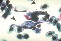
- In the macrophage, merozoites develop into megaloschizonts, which then divide into primary cytomeres. These replicate into smaller cytomeres, which in turn multiply through sporogony into merozoites. At this stage, merozoites will invade either white blood cells or developing red blood cells in order to become elongated gametocytes; these are usually 12-14 microns long.
- When a noninfected blackfly makes a blood meal on an infected bird, it ingests the elongated gametocytes. In the insect vector these elongated gametocytes become either a female (macro)gametocyte with a red-staining nucleus or a male (micro)gametocyte with a pale-staining diffuse nucleus, which come together to form an ookinete. This ookinete invades an intestinal cell of the fly vector, where it matures into an oocyst. This oocyst produces sporozoites which migrate to the salivary glands of the blackfly, thus reinitiating the cycle.
Ecology

Leucocytozoon's distribution depends on the habitat of its avian host species. L. simondi is suspected to be a major inhibitor of Canadian geese population growth in some areas, including the upper Midwestern United States and Canada. L. smithi affects turkey farms in the southeastern United States. Leucocytozoon does not threaten human populations in terms of potential infection; infected poultry is not pathogenic in humans. Still, the parasite's economic impact could hurt poultry farmers' revenue as mortality rates are extremely high, especially among young birds. Control of the blackfly vector is most likely the best way to cut down infections, but as blackflies reproduce rapidly and easily given the right conditions.
References
Butlet, J.F. Black flies, Simulium spp. University of Florida. 1998.<
Leucocytozoonosis. Michigan.gov. Department of Natural Resources.
Nahm, Julie et al. Leucocytozoon spp. University of Missouri College of Veterinary Medicine. 1997.
