Mammoth Cave
Overview
By Matt Selensky
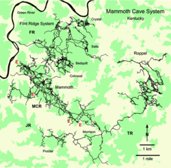
Caves around the world harbor myriad microbiota that thrive in these dark, energy-starved subsurface environments. Located in central Kentucky, Mammoth Cave is the longest known cave system on Earth, encompassing over 580 km of mapped passages[1]. Although a cave-wide assessment of its microbial ecology has never been performed, site-specific studies have elucidated intriguing characteristics of some Mammoth Cave microbes. Microbes found in the karstic sediments beneath two shallow water pools within Mammoth Cave were inferred to exhibit high diversity and total cell densities, reaching 1.4 × 10⁷ cells per g wet sediment[2]. Densities and activity of the chemolithoautotrophic Nitrobacter sp. were determined to be significantly higher in the caves relative to the surface. These nitrifying bacteria have been suggested to play a role in the formation of widespread saltpeter (Ca(NO3)2) deposits found in the cave by oxidizing bat guano N or other surface-sourced N transported underground[3]. A seep rich in hydrocarbons and sulfide in Marianne’s Pass brings additional energy sources into the cave for microorganisms[4]. No published work appears to be available that describes sulfur-oxidizing bacteria in Mammoth Cave. However, a 16S rRNA gene clone survey of nearby Parker Cave demonstrates the widespread abundance of Thiothrix sp. living in the underground and euxinic Sulphur River[5]. Caves untouched by humans inherently lack light; however, artificial lamps placed in “show cave” sections of Mammoth sustain photosynthetic algal and cyanobacterial populations[1]. Such microorganisms are otherwise thought to be transient, washing into the cave either after heavy rains or via riverine input[6]. Fungi commonly colonize and decompose organic matter such as rat fecal pellets or dead crickets that occasionally litter the cave passages. The most notorious fungus that is found in Mammoth Cave is Pseudogymnoascus destructans, the causative agent of white nose syndrome in bats[6]. Other eukaryotes that are found in the cave include amoebas and other “protists” that tend to colonize standing pools of water[6].
Geology of Mammoth Cave


Mammoth Cave is located in central Kentucky and is the longest known cave system on Earth, encompassing over 580 km of mapped passages [1]. Carved by dissolutional processes, the Mammoth Cave region is an example of a karst landscape. Sinkholes drain surface water into soluble limestone strata, which dissolve over geologic time. Mammoth Cave cuts through three major limestone layers, which were deposited 340-330 million years ago during the Mississippian Epoch[7]. The St. Louis limestone is the oldest of the three; it is dolomitic, 53-60 m thick, and commonly contains shale and chert nodules. The Ste. Genevieve limestone is 35 m thick and is somewhat less dolomitic than the St. Louis stratum, though shales are still common in this layer. The Girkin limestone is the youngest, least magnesium-rich carbonate layer in the Mammoth Cave region and is 40 m thick on average[7].
Although the limestones of Mammoth Cave are hundreds of millions of years old, the passages themselves are much younger[3][7]. An impermeable sandstone caprock exists over much of the region, which compacted and protected the underlying limestone from dissolution for millions of years. The caves began forming about 30 million years ago once the Green River eroded through the sandstone cap[3]. The formation of the caves (speleogenesis) occurred due to the flow of mildly acidic water through bedding planes[7]. Water becomes slightly acidic when it picks up CO2 from the atmosphere or overlying soils, where respiration produces abundant CO2. In water, CO2 exists in chemical equilibrium with carbonic acid (H2CO3); thus, as the partial pressure of CO2 increases, more H2CO3 is present in solution. The flow of this weak acid through pre-existing cracks and bedding planes dissolves the surrounding limestone, producing large passages over time. Following this paradigm, meteoric water tends to enter the Mammoth Cave system through numerous sinkholes in the Pennyroyal Plateau[7]. The water flows through the subsurface limestone until it discharges into the Green River, which continues to erode through the limestone layers.
As a limestone cave system, Mammoth Cave is dominated by carbonate geochemistry, yielding alkaline-buffered solutions. However, some parts of the cave system exhibit distinct geochemical characteristics. Beneath the Mammoth Cave region lies a monocline, which cuts through most of the South-Central Kentucky Karst[4]. There are several sites along this monocline where sulfide-laden brines rise to the surface, including Sulphur River in nearby Parker Cave[4][5]. Marianne’s Pass within Mammoth Cave also receives sulfide-rich brines, though the origins of this seep are likely not from the same monocline structure[4].
Microbial Ecology of Mammoth Cave
Like most cave environments, the microbial ecology of Mammoth Cave is just beginning to be understood. In this dark habitat, photosynthesis is impossible, save for isolated areas around installed lamps in tourist caves. As such, microorganisms living within caves are either dependent on organic carbon and nutrients washed in from the surface, or by organic carbon fixed within the caves themselves by chemolithoautotrophs.
A cave-wide, broad taxonomic survey of Mammoth Cave microbiota has yet to be performed. Nonetheless, several studies have characterized specific microorganisms colonizing certain environments within this and nearby cave systems[1][2][3][5].
One such study investigated bacterial abundance, activity, and diversity in waterlogged sandy sediments 100m below the surface [2]. These sediments were located within the Echo River basin and are fed by water percolating from the ceiling. Total microbial abundance was assessed by counting cells via fluorescence microscopy using acridine orange (AO). By pre-staining cells with p-iodonitrotetrazolium violet (INT) four hours before the AO stain, the abundances of viable cells were determined in these sediments, since only living cells incorporate INT. Cell densities were similar between the two sediments, reaching 1.4 × 10⁷ cells per g wet sediment, with over 50% of all cells determined to be metabolically active based on INT incorporation[2].
The microbial diversity of these same sediments was evaluated by an agar plate-based cultivation approach[2]. Agar plates were prepared by extracting sediment with DI water to mimic in situ conditions. These plates were then inoculated with fresh cave sediment. After isolating individual colonies from these plates, isolated strains were identified by examining their colony morphology and biochemical characteristics. Using this approach, 44 individual strains were identified, representing members of the Alcaligenes, Staphylococcus, Chromobacterium, Mycobacterium, Flavobacterium, Corynebacterium, Aureobacterium, Arthrobacter, Brevibacterium, Actinobacteria, Bacillus, Micrococcus, and Curtobacterium. Many of the cultured isolates were able to utilize cellulose or its breakdown products as a sole carbon source[2]. Since these organic compounds are very resistant to degradation, it is assumed that these strains must rely on compounds that surface microorganisms do not consume[2]. It is important to note, however, that chemolithoautotrophic activity in the cave sediments was not assessed by these methods; such carbon fixing microbes may produce labile organic carbon in situ that could have been missed by this survey. Nevertheless, this was among the first studies to recognize that microorganisms within Mammoth Cave sediments “significantly affect nutrient cycling, the degradation of complex compounds, and carbon flow”[2].

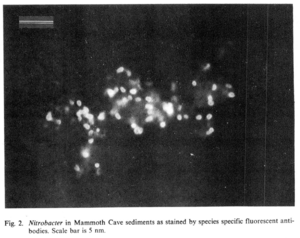
The activity of chemolithoautotrophs throughout Mammoth Cave has yet to be evaluated, though there is reason to believe they exist and are active. Only one study has directly examined the presence of these carbon-fixing microbes in the caves[3]. Using a fluorescent antibody-based approach, Fliermans & Schmidt (1977) determined that the cell densities of chemolithoautotrophic Nitrobacter sp. in Mammoth Cave sediments were 6.2 × 10⁵ cells per g compared to 103 cells per g of surface soils. Of the Mammoth Cave Nitrobacter population, 85% were identified as N. agilis. As nitrifying bacteria, Nitrobacter sp. oxidize nitrite to nitrate as part of their energy metabolism. The origins of nitrite in the caves are unknown, with explanations ranging from surface runoff to the degradation of bat guano[3]. Nitrogen is a limiting nutrient in oligotrophic environments such as caves; as such, it is intriguing that taxa capable of nitrification are enriched in Mammoth Cave. The presence of other nitrogen cycling microbes in Mammoth Cave, such as diazotrophs or denitrifiers, is unknown. See below for further discussion on the presence of Nitrobacter in Mammoth Cave.
At least one sulfur-rich hydrocarbon seep has been described in Mammoth Cave[4]. This seep is located within Marianne’s Pass and is not thought to be sourced from the same brines that are associated with the regional-scale monocline structure[4]. Although the origin of the fluids discharging in Marianne’s Pass is unknown, the sulfide in this seep provides a source of electrons that chemolithoautotrophs could exploit within Mammoth Cave. No published studies investigate the microbial ecology of the Marianne’s Pass seep, but this is an active area of investigation.
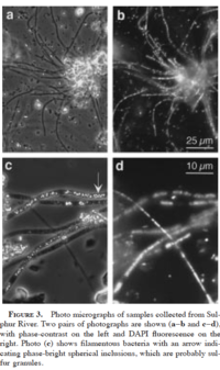
In fact, the microbial ecology of Mammoth Cave has not been assessed by culture-independent approaches such as environmental DNA sequencing in a peer-reviewed publication. However, environmental DNA has been sequenced from a subterranean river in Parker Cave, located nearby[5]. The underground Sulphur River discharges within Parker Cave. One of its tributaries, Phantom Waterfall, is euxinic. The mud floor surrounding the Phantom Waterfall has a pH of 0.13 and contains visible deposits of elemental sulfur. Phantom Waterfall is thought to be composed of rainfall-derived groundwater mixed with natural hydrocarbons. The ceiling is coated with an acidic slime. Due to carbonate buffering from the limestone walls of the cave, the pH of the downstream Sulphur River is 6.4 to 7.6. A 16S rRNA gene survey of white biofilms at the confluence of the Phantom Waterfall and Sulphur River showed the predominance of sulfate-reducing and sulfide-oxidizing bacteria in this sulfide-laden habitat[5]. Some of the 16S rRNA gene sequences were assigned to chemolithoautotrophic sulfide oxidizer Thiothrix ramosa[5]. These data suggest a microbially-driven S cycle that could support C fixation via chemolithoautotrophy within this dark habitat. Although this analysis was performed in a nearby cave, its implications likely also apply to some portions of Mammoth Cave, such as Marianne’s Pass.
Bacteria are not the only microbes that exist within Mammoth Cave; descriptions of cave eukaryotes also exist. Protists have been found inhabiting cave waters, guano, and sediments[6]. In general, waters that are directly connected to the surface exhibit higher cell densities of micro-eukaryotes compared to more isolated cave pools[6]. Fungi are commonly found throughout Mammoth Cave, colonizing bat guano, rat excrement, and other organic carbon sources imported from the surface[6]. The most notorious fungus that calls Mammoth Cave home is Pseudogymnoascus destructans, the causative agent of White Nose Syndrome (WNS) in bats[6][8]. See "A Deadly Fungus" for further discussion pertaining to WNS.
To date, no studies have been published that describe Archaea in Mammoth Cave.
Guns, Germs, and Guano
Some sections of Mammoth Cave have long been a productive source of raw saltpeter (calcium nitrate, Ca(NO3)2), one of the core components of gunpowder. Cave sediments rich in Ca(NO3)2 were extensively mined to produce gunpowder; over 1800 tons of sediment were extracted from Mammoth Cave during the War of 1812 alone[3]. Despite the importance of these nitrate-rich sediments for military purposes, their origins remain elusive.

Early theories postulated that nitrates deposit in the cave after descending water leaches nitrate from sandstone, which can commonly contain NO3-[3]. This is consistent with the geological setting of Mammoth Cave, which underlies sandstone caprocks[7]. However, the prevailing theory of saltpeter formation at the time invoked the breakdown of nitrogen-rich bat guano as described by references in [3]. Nonetheless, sediments rich in saltpeter are found much deeper in Mammoth Cave than bats generally travel[3].
Alternatively, chemolithoautotrophic nitrifying bacteria could be the main source of nitrates in Mammoth Cave sediments[3]. In 1977, Fliermans & Schmidt were the first to directly investigate this possibility. Before the advent of widespread environmental DNA sequencing techniques, they employed a culture-independent fluorescent antibody method to quantify the population sizes of Nitrobacter agilis and N. winogradskyi in Mammoth Cave sediments compared to overlying soils[3].
To accomplish this, Fliermans & Schmidt (1977) first grew pure cultures of N. agilis and N. winogradskyi in the laboratory. Aliquots of these pure cultures were then introduced to carrier rabbits, which produced antibodies against the specific Nitrobacter species of interest. The antibodies were then harvested, isolated, and conjugated to different fluorescent compounds, depending on the Nitrobacter sp. The subsequently produced fluorescent antibody (FA) acted as a stain for fluorescent microscopy. Cells from the environment could then be counted under the microscope, with the two Nitrobacter sp. glowing different colors.
As discussed above, the abundance of Nitrobacter sp. in the cave sediments were over two orders of magnitude higher than surface soils[3]. Additionally, cells were counted before and after water was used to extract nitrate from sediment samples in nitrate leaching experiments. The abundance of Nitrobacter cells did not change after water was used to extract nitrate from sediment samples [3]. Field observations noted that [NO3-] returns to the concentrations measured before leaching within a few years[3]. This, along with the independence of these cells from NO3-, was taken as evidence that cave Nitrobacter, a known nitrite oxidizer, produces nitrate. Despite this conclusion, the source of the more reduced forms of nitrogen remains unknown in these caves.
Photosynthesis in Mammoth Cave
Some tourist sections of Mammoth Cave are artificially lit with lamps, which provides a source of photons for photosynthetic mosses, ferns, algae, and cyanobacteria, which could otherwise not survive in the dark cave habitat[1]. A survey of these so-called “lamp florae” identified 28 invasive photosynthesizers within Mammoth Cave[1]. Of these 28 species, 14 were classified as cyanobacteria, 8 were algae, and 6 were diatoms. This is a concern for park officials, as these lamp florae can quickly displace native microflora and can damage delicate cave features. An increase in biomass on the walls of these passages results in higher rates of respiration; the concomitantly produced carbonic acid can enhance the dissolution of limestone features[1].
Mitigating the spread of lamp florae has been a major focus of Mammoth Cave management since the installation of lamps in the early 1940s[9]. Traditionally, applying household bleach, formaldehyde, and hydrogen peroxide solutions to infested cave walls has been used to remove or slow the growth of invasive flora[9]. However, these chemical treatments can pose safety hazards and are equally devastating to native cave microbes. To overcome these issues, Olson 2006 proposed to prevent the growth of photosynthesizers by changing the lamps themselves. Cave lamps traditionally contain incandescent or halogen bulbs, which emit light across the visible electromagnetic spectrum. In infested portions of the Frozen Niagara section in Mammoth Cave, these lamps were replaced with yellow LEDs[9]. These lamps emit a narrower wavelength spectrum, peaking at 595 nm. Since chlorophyll pigments absorb light most strongly in the 400-475 and 600-700 nm ranges, it was hypothesized the installation of yellow LEDs would inhibit growth by not providing those optimal wavelengths[9]. After 2.5 years of use in Frozen Niagara, cave sections illuminated with the yellow LEDs did not harbor invasive lamp flora[9].
A deadly fungus: P. destructans and white nose syndrome

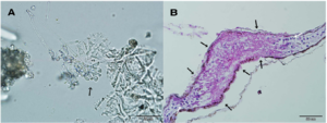
White Nose Syndrome (WNS) is a deadly bat disease caused by the fungus Pseudogymnoascus destructans, formerly Geomyces destructans[8][6]. P. destructans is native to Europe but was introduced to the United States in 2006[6]. Since then, over 6.7 million bats in North America have succumbed to the disease[10], which infects the skin of bats and disrupts hibernation[8].
During hibernation, bat body temperature decreases to conserve metabolic energy in the winter months. Bats generally undergo periods of torpor between 12.4 and 19.7 days interrupted by short periods of arousal for 1-2 hours[10]. In these arousal periods, bat metabolic rate increases dramatically; in fact, 80-90% of the energy used by bats in the winter months is consumed during intermittent arousal[10]. P. destructans infects the skin of bats, irritating it and possibly causing water loss to the tissues[8][10]. This results in bats awakening from their torpor more frequently, causing them to deplete their energy reserves stored as fat[10]. In this way, WNS can decimate bat populations, with mortality rates commonly exceeding 90% in some species of North American bat[10]. Managing the spread of P. destructans is a major concern for scientific and recreational caving alike[6].
Key Microbial Players
Conclusion
Cave microbiota are relatively understudied and represent a hidden cache of microbial diversity and function in the subsurface. Despite a general lack of cave-wide taxonomic surveys, site-specific studies demonstrate that the microbial communities of Mammoth and other caves are greatly involved in the N, S, and C cycles. We are only just beginning to gain a system-level understanding of shallow subsurface microbial ecology.
References
- ↑ 1.0 1.1 1.2 1.3 1.4 1.5 1.6 Smith T, and Olson RA. (2007) A taxonomic survey of lamp flora (algae and cyanobacteria) in electrically lit passages within Mammoth Cave National Park, Kentucky. International Journal of Speleology, 36: 105-114.
- ↑ 2.0 2.1 2.2 2.3 2.4 2.5 2.6 2.7 Rusterholtz KJ, and Mallory, LM. (1994) Density, activity, and diversity of bacteria indigenous to a karstic aquifer. Microbial Ecology, 28:79-99.
- ↑ 3.00 3.01 3.02 3.03 3.04 3.05 3.06 3.07 3.08 3.09 3.10 3.11 3.12 3.13 3.14 3.15 Fliermans CB, and Schmidt EL. (1977) Nitrobacter in Mammoth Cave. International Journal of Speleology, 9: 1-19.
- ↑ 4.0 4.1 4.2 4.3 4.4 4.5 Olson R. (2013). Potential effects of hydrogen sulfide and hydrocarbon seeps on Mammoth Cave ecosystems. Mammoth Cave Research Symposia, 28: 25-31.
- ↑ 5.0 5.1 5.2 5.3 5.4 5.5 5.6 Angert, ER, Northup, DE, Reysenbach, AL, Peek, AS, Goebel, BM, and Pace, NR. (1998) Molecular phylogenetic analysis of a bacterial community in Sulphur River, Parker Cave, Kentucky. American Mineralogist, 83: 1583-1592.
- ↑ 6.0 6.1 6.2 6.3 6.4 6.5 6.6 6.7 6.8 6.9 Lavoie, KH (2017) Mammoth Cave Microbiology. In: Mammoth Cave: A Human and Natural History. edited by HH Hobbs, RA Olson, EG Winkler, and DC Culvers, Springer International Publishing, Cham, pp 235-250.
- ↑ 7.0 7.1 7.2 7.3 7.4 7.5 7.6 7.7 Palmer, A. Mammoth Cave Geology. In: Mammoth Cave: A Human and Natural History. edited by HH Hobbs, RA Olson, EG Winkler, and DC Culvers, Springer International Publishing, Cham, pp 235-250.
- ↑ 8.0 8.1 8.2 8.3 8.4 Zukal, J, Bandouchova, H, Brichta, J, et al. (2016) White-nose syndrome without borders: Pseudogymnoascus destructans infection tolerated in Europe and Palearctic Asia but not in North America. Scientific Reports, 6, 19829.
- ↑ 9.0 9.1 9.2 9.3 9.4 Olson, RA. (2006) Control of lamp flora in developed caves. Cave conservation and restoration, Huntsville, National Speleology Society, 343-348.
- ↑ 10.0 10.1 10.2 10.3 10.4 10.5 Reeder, DM, Frank, CL, Turner, GG, et al. (2012) Frequent arousal from hibernation linked to severity of infection and mortality in bats with white nose syndrome. PLoS One, 7(6): e38920.
Authored for Earth 373 Microbial Ecology, taught by Magdalena Osburn, 2020, NU Earth Page.
