Microbiota of Patients with Atopic Dermatitis
Background Information

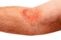
Atopic dermatitis, also known as eczema, is a chronic inflammation of the skin that affects about 17% of the population, mostly infants and young children [1]. Symptoms include itchiness, redness, dryness, and pain due to breaches in the skin. Breaches to the protective barrier of the skin allow bacteria to enter and establish an infection. The normal flora of the skin normally does not cause infections in healthy individuals. However, atopic dermatitis creates an exposed area on the skin for opportunistic pathogens to cause an infection. Studies have shown that the microbiota of patients suffering from atopic dermatitis differ from the microbiota of healthy people.
Skin Microbiome

Healthy Persons
The skin is the largest organ in the human body. It is inhabited by many bacteria that are usually harmless and serve to out-compete potentially harmful bacteria [3]. These commensal bacteria have high diversity which allows them to colonize different areas of the body. The surface of the skin such as the forearms, back of the hands, legs, and back can be dry and high in salt concentration. This can inhibit some species of bacteria that cannot grow in dry and salty environments but allow other species to thrive in this environment. On the other hand, moist areas on the skin such as between the toes, behind the knees, and under the arms are ideal environments for some bacteria.
Key Organisms
It was commonly thought that the key organisms inhabiting the human skin were "Staphylococcus epidermidis" and "Staphylococcus aureus" but research has shown that this is not the case. A research done by sequencing the 16S ribosomal RNA obtained from the skin of five healthy volunteers ranging in age, race, and ethnicity showed that only 5-10% of the skin microbiota was "S. epidermidis" or "S. aureus" [4]. The dominant inhabitants of the skin normal flora belong to the four phyla: Proteobacteria, Actinobacteria, Bacteroidetes, and Firmicutes in the order of abundance on the skin [4].
Patients with Atopic Dermatitis
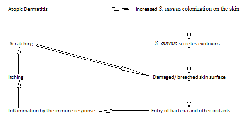
Patients with atopic dermatitis show defects in their innate and adaptive immune response which increases the risk of bacterial and viral infections [5]. Thus, patients with atopic dermatitis have more colonization of "S. aureus" on the skin, leading to complications of the skin disease [5] [6] [7]. "S. aureus" secretes exotoxins that aggravate inflammation by inducing an inflammatory immune response against the pathogen [7]. This leads to more itching and scratching of the infected area which elevates the symptoms of atopic dermatitis.
Gut Microbiome
Healthy Persons
There are over 1000 bacterial species that live in the gut of humans [8]. These anaerobic organisms help with the digestion of foods and absorption of nutrients in the intestines [9] [10]. Research shows that the microbiota of humans contribute about 360 times more genes than the human genome [10]. These microbial genes are necessary for human survival and can encode for enzymes to digest the food that would otherwise not be digested [10]. The diversity of the gut normal flora is established upon birth and further develops as food containing a variety of microorganisms is ingested.
Key Organisms
The key microorganisms present in the gut are categorized into two phyla: Firmicutes and Bacteroidetes [8]. Research shows that the balance between these two dominant phyla in the gut controls the energy metabolism of the host and imbalances can lead to metabolic diseases [8].
Patients with Atopic Dermatitis
Infants that have atopic dermatitis have a lower diversity of bacteria in the gut compared to healthy infants [11]. The relative abundance of microbes from different phyla in infants with atopic dermatitis was determined and compared with healthy infants and discovered that there was a lower abundance of Bacteroidetes and Proteobacteria in infants with atopic dermatitis [11]. With a less variety of microorganisms exposed to the gut at infancy when the immune system is maturing, the prevalence of developing allergic diseases such as atopic dermatitis increases due to changes in the immune modulation [12].
Treatments
The basic treatment of atopic dermatitis is skin care [13]. This includes moisturizing the skin, avoiding non-specific irritants such as wool, and avoiding irritating factors such as soap, hot water, extreme pH solutions, and allergens [13]. Further treatments are also available depending on the severity of the disease [13]. Examples of further treatments include emollients, corticosteroids, antimicrobial therapy, and UV therapy [13].
References
[1] Laughter D, Istvan JA, Tofte SJ, Hanifin JM. “The prevalence of atopic dermatitis in Oregon school children.” Journal of the American Academy of Dermatology, 2000, 43: 649-655.
[2] Vivacare Inc. “Atopic Dermatitis.” From Your Doctor. (2013). < http://fromyourdoctor.com/Condition_Centers/Eczema_Basics_Center/Category/Atopic_Dermatitis>
[3] Chiller K, Selkin BA, Murakawa GJ. “Skin Microflora and Bacterial Infections of the Skin.” Journal of Investigative Dermatology Symposium Proceedings, 2001, 6: 170–174.
[4] Grice EA, Kong HH, Renaud G, Young AC, NISC Comparative Sequencing Program, Bouffard GG, Blakesley RW, Wolfsberg TG, Turner ML, Segre JA. “A diversity profile of the human skin microbiota.” Genome Research, 2008, 18(7):1043-50.
[5] Baker BS. “The role of microorganisms in atopic dermatitis.” Clinical & Experimental Immunology, 2006, 144(1): 1-9.
[6] Kong, HH, Oh J, Deming C, Conlan S, Grice EA, Beatson MA, Nomicos E, Polley EC, Komarow HD, NISC Comparative Sequence Program, Murray PR, Turner ML, Segre JA. “Temporal shifts in the skin microbiome associated with disease flares and treatment in children with atopic dermatitis.” Genome Research, 2012, 22: 850–859.
[7] Yu TL, Chen TW, Bor LC. “Role of Bacterial Pathogens in Atopic Dermatitis.” Clinical Reviews in Allergy & Immunology, 2007, 33(3): 167-177.
[8] Burcelin R, Luche E, Serino M, Amar J. "The gut microbiota ecology: a new opportunity for the treatment of metabolic diseases." Front Biosci, 2009, 14(5): 107-5117.
[9] McFarland LV. “Normal flora: diversity and functions.” Microbial Ecology in Health and Disease, 2000, 12: 193–207.
[10] “NIH Human Microbiome Project defines normal bacterial makeup of the body.” National Institutes of Health, 2013, <http://www.nih.gov/news/health/jun2012/nhgri-13.htm>
[11] Abrahamsson TR, Jakobsson HE, Andersson AF, Björkstén B, Engstrand L, Jenmalm MC. “Low diversity of the gut microbiota in infants with atopic eczema.” The Journal of Allergy and Clinical Immunology, 2012, 129(2): 434-440.
[12] Cox MJ, Huang YJ, Fujimura KE, Liu JT, McKean M, Boushey HA, Segal MR, Brodie EL, Cabana MD, Lynch SV. “Lactobacillus casei Abundance Is Associated with Profound Shifts in the Infant Gut Microbiome.” PLoS ONE, 2010, 5(1): e8745. doi:10.1371/journal.pone.0008745
[13] Akdis CA, Akdis M, Bieber T, Bindslev-Jensen C, Boguniewicz M, Eigenmann P, Hamid Q, Kapp A, Leung DYM, Lipozencic J, Luger TA, Muraro A, Novak N, Platts-Mills TAE, Rosenwasser L, Scheynius A, Simons FER, Spergel J, Turjanmaa K, Wahn U, Weidinger S, Werfel T, Zuberbier T, the European Academy of Allergology and Clinical Immunology/American Academy of Allergy, Asthma and Immunology/PRACTALL Consensus Group, “Diagnosis and treatment of atopic dermatitis in children and adults: European Academy of Allergology and Clinical Immunology/American Academy of Allergy, Asthma and Immunology/PRACTALL Consensus Report.” Allergy, 2006, 61: 969–987.

