Nocardia
Classification
Domain: Bacteria Phylum: Actinobacteria Class: Actinobacteria Order: Actinomycetales Family: Nocardiaceae Genus: Nocardia Species: N. asteroides, N. brasiliensis, N. farcinica, N. veterana
Description
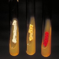
The genus Nocardia was first discovered by Edmund Nocard, a French veterinarian, in 1889, as the main cause of bovine infections [1]. The species of Nocardia genus are gram-positive and rod-shaped bacteria. Different species can range have colony colors from white, to tan, orange and even red in color. Colonies appear to be smooth or moist, and can have a "mold-like" appearance. These microbes are no more than 2 microbes in thickness and length. They are reliant on getting their nourishment from dead and/or decaying organic material, making these bacteria saprophytic. Nocardia are commonly found in soils, plants, animal tissues, and human tissues [2].
The bacteria in this species have been common to diseases. Nocardiosis, the most common disease associated with these genus, which is an infection disease affecting either the lungs or the entire body. Also, the inhalation of Nocardia species can lead to pneumonia, as well as skin infections if they enter through open wounds [3].
Nocardia species have been closely related to species from the genus Rhodococcus. There are 14 hypothetical conserved proteins that are unique to Nocardia and Rhodococcus. [4]

Left tube: Actinomadura madurae, Middle tube: Nocardia asteroides, Right tube: Micromonspora.
Image provided by CDC/Dr. David Berd
Ecology and Significance
Nocardiagrow slowly on nonselective culture media, and are strict aerobes that can grow at a wide range of temperatures. These bacteria can be cultured and colonies become evident in about 3-5 days, but sometimes a prolonged incubation period of 2-3 weeks is necessary to see colony formation. They grow on carbon rich soils, plants, and animals. Once it lands on its host, it immediately begins metabolizing tissues for energy or nutrients. [5]
Nocardia related infections have a high mortality rate and are resistant to treatment. They are particularly harmful in patients that have suppressed immune systems, similar to those with HIV or patients that have recently gone through a transplant procedure. Some drugs that currently show any effect against this genus are ketoconazole, clotrimazole, and miconazole. [6]
This genus was only sequenced three years ago, so it seems reasonable that there has not been any clear evidence of significance just yet. As research continues that may change.
Genome Structure
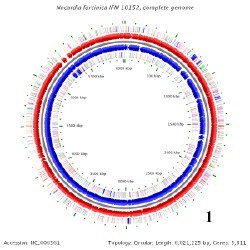
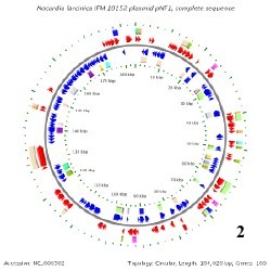
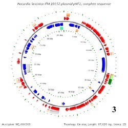
There is only one known genome in the Nocardia species and that belongs to Nocardia farcinica. This genome contains a circular chromosome that is 6,021,225 base pairs long, containing 5,811 genes, and having a GC content around 71%. N. farcinica has two plasmids that go along with its chromosome, pNF1 and pNF2. pNF1 is 184,027 base pairs long, consisting of 160 genes, and with a GC content of 67%. pNF2 is 87,093 base pairs long, made up of 93 genes, and has a GC content of 60%. All Of these information pertaining to the genome have been determined from 16S rRNA sequencing. [7]
Below is BacMap Genome Mapping, courtesy of Stothard P, Van Domselaar G, Shrivastava S, Guo A, O'Neill B, Cruz J, Ellison M, Wishart DS (2005) BacMap: an interactive picture atlas of annotated bacterial genomes. Nucleic Acids Res 33:D317-D320.
 ****
**** ****
****
Image 1: N. farcinica Chromosome
Image 2: pNF1 (Plasmid 1)
Image 3: pNF2 (Plasmid 2)
Metabolism
Species in the genus Nocardia carry out a broad variety of metabolic pathways. Their carbohydrate metabolism includes glycolysis, TCA cycle, pentose phosphate, and an extremely long list of sugar metabolic pathways. One species, N. farcinica, actually carries out two photosynthetic pathways. Those being the carbon fixation pathway and reductive carboxylate cycle.
A secondary metabolism has been studied, and describes the high quantity of oxygenases found in the genus. It proposes that the purpose might be for fatty acid metabolism, or a defense system against toxins. [8]
References
[1] Jun Ishikawa, Atsushi Yamashita, Yuzuru Mikami, Yasutaka Hoshino, Haruyo Kurita, Kunimoto Hotta, Tadayoshi Shiba, and Masahira Hattori. The complete genomic sequence of Nocardia farcinica IFM 10152. 101:41 (2004): Web.
[2] Segawa, Shunsuke. "Identification of Nocardia species using matrix-assisted laser desorption/ionization-time-of-flight-mass spectrometry." 12:6 (2015): Web.
[3] Stothard P, Van Domselaar G, Shrivastava S, Guo A, O'Neill B, Cruz J, Ellison M, Wishart DS (2005) BacMap: an interactive picture atlas of annotated bacterial genomes. Nucleic Acids Res 33:D317-D320
[4] Munoz J, Mirelis B, et al., Clinical and microbiological features of nocardiosis 1997-2003.
[5] Edward P. Desmond, Martha Flores. Mouse pathogenicity studies of N. asteroides complex species and clinical correlation with human isolates. Volume 110 Issue 3 Page 281Issue 3 - 284 - July 1993.
[6] Gao, Beile, and Radhey S. Gupta. “Phylogenetic Framework and Molecular Signatures for the Main Clades of the Phylum Actinobacteria.” Microbiology and Molecular Biology Reviews : MMBR 76.1 (2012): 66–112. PMC. Web.
[7] M M McNeil and J M Brown. The medically important aerobic actinomycetes: epidemiology and microbiology. Clin Microbiol Rev. 1994 July; 7(3): 357–417.
[8] http://en.wikipedia.org/wiki/Nocardia#Culture_and_staining
Author
Page authored by Alex Jenkins, student of Prof. Katherine Mcmahon at University of Wisconsin - Madison.