Photobacterium leiognathi
A Microbial Biorealm page on the genus Photobacterium leiognathi
Classification
Higher order taxa
Domain (Bacteria); Phylum (Proteobacteria); Class (Gammaproteobacteria); Order (Vibrionales); Family (Vibrionales); Genus (Photobacterium)
Species
|
NCBI: Taxonomy |
Photobacterium leiognathi
Description and significance
Many have been mystified by the beauty of the “glowing” seas. Bioluminescent bays are rare and fragile ecosystems. Photobacterium leiognathi is amongst the organisms that help provide its natural glow.
P. leiognathi is one of the many species within the genus Photobacterium[1] and the family of Vibrionales. P. leiognathi is a gram-negative, coccobacillus-straight shaped, flagellated bacterium. P. leiognathi has no pigmentation and appears white or colorless unless found in colonies where it exhibits luminescent properties. P. leiognathi is a facultative anaerobic chemoorganothrophic bacterium. P. leiognathi lives as a free-living organism in temperate marine water or can be found in association with fish. [8,10,11,12]
Genome structure
Like many other Photobacteria P. leiognathi genetic material is found in two circular chromosomes. Currently the highest level of assembly is scaffold only with 4,598,918 total bases with nucleotide sequence of 7.3 kbp. A key component of its genome is the lux-rib operon which includes the following genes: luxR (repressor), luxI (inducer), luxA, luxB (luciferase), luxC, luxD, luxE (aldehyde), and luxG (not known) which are responsible for the bioluminescence properties of the bacteria. It is worth noting that P. leiognathi lacks the luxF gene whose functions and role are not clearly identified yet. Due to the sequencing of the 16S rRNA, many species, including this one, have occasionally been shifted in and out of the genus, from genus Photobacterium to genus Vibrio. Some genomic polymorphism has been seen within the species depending on the type of host they form a symbiotic relationship with. Currently 11 subspecies of P. leiognathi are known. [11, 12, 7, 3, 10, 1, 4, 9, 10, 13]
Cell and colony structure
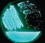
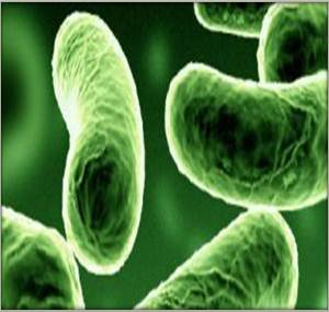
P. leiognathi is a gram negative bacterium with coccobacillus shaped cells. The cells can be straight or plump rods. The rods have a diameter of about ~0.8 - 1.6 µm and a length of ~1.8 - 2.4 µm. P. leiognathi can contain 1-3 polar flagella, but some cells found in symbiotic relationship with fish have shown to be non-motile. The individual cells contain no pigmentation and appear white or colorless. Interestingly, when the bacterium forms colonies they exhibit luminescent properties, emitting bluish-green light of ~490nm. Luminescence is only exhibited when the bacteria is found in colonies because a high concentration of an autoinducer released by the bacteria is necessary to activate the lux gene. It is believed that P. leiognathi, like many other Photobacteria, utilizes the quorum sensing mechanism to detect the right concentration of the auto-inducing enzymes and trigger luciferase production. Luciferase is then responsible for controlling the oxidation reaction of luciferin in which riboflavin mononucleotide (FMNH2) and a long chain aldehyde are reduced. The reaction produces luminescence, a continuous glow. [1, 7, 11, 8, 12, 13]
Metabolism
P. leiognathi is a marine, organism, therefore it requires sodium for growth. P. leiognathi is a chemoorganotrophic[2] microorganism that obtains its energy from the oxidation of organic compounds like glucose, mannose, fructose, or glycerol. P. leiognathi is a facultative anaerobe [3] which means it can perform both fermentation in the absence of oxygen and respiratory metabolic functions in the presence of oxygen. Like many other Photobacteria, P. leiognathi prefers an environment with oxygen, because oxygen plays a key role in the production of luminescence [4] [11, 13]
Ecology
P. leiognathi might be found as free-living organism in warm tropical marine environments within the mesopelagic zone (200-1000m depth) or in association with fish. P. leiognathi grows best in water at temperature between 18-30 °C. P. leiognathi most studied association appears to be a symbiotic relationship with the ponyfish. In fact, the ponyfish has developed a special compartment, light organ, where the bacteria can live, thrive and survive. P. leiognathi might also be found as a neutral entity on the surface of fish or acting as a decomposer of dead fish. [2, 3, 9, 10, 11]
Pathology
Other species of the genus Photobacterium have been found to be pathogenic to marine life and mammals, but not P. leiognathi. The transmembrane DNA binding protein ToxR and the associated membrane protein ToxS can be found in many ocean species that are pathogenic to human or fish. P. leiognathi contains ToxR, however no evidence of pathogenicity has been found in the species. [7, 12]
References
[1] Nijvipakul, Sarayut, et al. 2008, "LuxG Is a Functioning Flavin Reductase for Bacterial Luminescence", American Society for Microbiology: Journal of Bacteriology, Vol. 190, No. 5, 1531-1538,doi: 10.1128/JB.01660-07
[2] Valentine N. Petushkov, Bruce G. Gibson,and John Lee. 1995, “Properties of recombinant fluorescent proteins from Photobacterium leiognathi and their interaction with luciferase intermediates”, Biochemistry including biophysical chemistry and molecular biology, Vol. 34, No. 10, 3300-3309, DOI: 10.1021/bi00010a020
[3] Lee,Chan Yong, Rose B. Szittner, and Edward A. Meighen. 1991, “The lux genes of the luminous bacterial symbiont, Photobacterium leiognathi, of the ponyfish; nucleotide sequence, difference in gene organization, and high expression in mutant Escherichia coli”, European Journal of Biochemistry, Vol. 201, No. 1, 161-167, DOI: 10.1111/j.1432-1033.1991.tb16269.x
[4] Herring, Peter. 2002, “Marine microlights: the luminous marine bacteria” Microbiology Today, Vol. 29, 174-176
[5] Coil, David. 2011, “Microbiology Christmas Tree – luminescent bacteria,giant microbes, and more”, Microbiology of the Built Environment Network
[6] Boisvert, H. 1967, “Photobacterium leiognathi”, National Center for Biotechnology Information
[7] Yirka, Bob. 2011, “Research shows ocean bacteria glow to attract those that would eat them” Physorg.com. doi: 10.1073/pnas.1116683109
[8] Haddock, Steven H.D., Mark A. Moline, and James F. Case. 2010 “Bioluminescencein the Sea”, Annual Review of Marine Science, Vol. 2, 443-493
[9] Malave-Orengo, J., Eva N. Rubio-Marrero, and C. Rios-Velazquez. 2010 “Isolation and characterization of bioluminescent bacteria from marine environments of Puerto Rico”, Formatex, Vol. 2, 103-108
[10] Medvedeva, S. E., O. A. Mogil’naya, and L. Yu. Popova. 2006, “Heterogeneity of the populations of marine luminescent bacteria Photobacterium leiognathi under different conditions of cultivation”, Microbiology, Vol. 75, No. 3, 292-295, DOI: 10.1134/S002626170603009X
[11] Herring, P. J. and Widder, E. A., 2001 "Bioluminescence in Plankton and Nekton", Encyclopedia of Ocean Science, Vol. 1, 308-317
[12] Dunlap, Paul V., et al. 2004 “Genomic polymorphism in symbiotic populations of Photobacterium leiognathi”, Environmental Microbiology, Vol. 6, No. 2, 145-158, DOI: 10.1046/j.1462-2920.2003.00548.x
[13] Fuqua, W. C., S.C. Winas, and E.P. Greenberg. 1994. “Quorum sensing in bacteria: the LuxR-LuxI family of cell density-responsive transcriptional regulators”, Journal of Bacteriology, Vol. 176, No. 2, 269–275
Edited by Jossary Gerry, student of Dr. Lisa R. Moore, University of Southern Maine


