Propionibacterium
A Microbial Biorealm page on the genus Propionibacterium
Classification
Higher order taxa:
Bacteria; Actinobacteria; Actinobacteria (class); Actinobacteridae; Actinomycetales; Propionibacterineae; Propionibacteriaceae
Species:
Propionibacterium acidipropionici; P. acnes; P. australiense; P. avidum; P. cyclohexanicum; P. freudenreichii; P. granulosum; P. jensenii; P. microaerophilum; P. propionicum; P. thoenii; P. sp.
|
NCBI: Taxonomy Genome: -Nocardioides sp. JS614 -Propionibacterium acnes KPA171202 |
Description and Significance
Species of Propionibacteria can be found all over the body. Propionibacteria are generally nonpathogenic; however, when certain species of Propionibacteria contaminate blood and other body fluid, they can cause a number of infections including the common skin disease acne vulgaris (caused by P. acnes). Some species of Propionibacteria are found in food like cheeses and other dairy products. P. freudenreichii is used in Swiss cheese manufacturing to produce its flavor and characteristic holes. The flavor derives from propionate, a product of fermentation, and the holes or "eyes" from bubbles of carbon dioxide.
Genome Structure
Currently there is one genome project that is complete on Propionibacterium acnes KPA1202, and there is one is progress on Nocardioides sp. JS614. The entire genome sequence of P. acnes includes 2333 putative genes and reveals numerous gene products involved in degrading host molecules, including sialidases, neuraminidases, endoglycoceramidases, lipases, and pore-forming factors. For more information see links (above, left).
Cell Structure and Metabolism
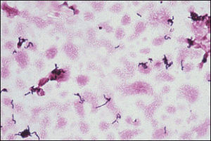
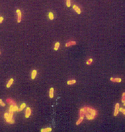
Propionibacteria are slow-growing, nonsporeforming, Gram-positive, anaerobic bacteria. They can be rod-shaped or branched and can occur singularly, in pairs, or in groups. They generally produce lactic acid, propionic acid, and acetic acid from glucose.
One species of Propionibacterium, P. freudenreichii (used in Swiss cheese manufacturing), seems to be dependent on free amino acids and peptides made in the cheese by casein degradation of Lactobacillus proteolytic enzymes (McCarthy and Courtney). P. freudenreichii produces carbon dioxide, which makes the holes in swiss cheese. The propionic acid produced from breaking down glucose is what gives Swiss cheese its characteristic flavor.
Ecology
Although Propionibacterium acnes can be found on the skin of prepubesent humans, true colonization actually begins 1 to 3 years before sexual maturation. The numbers of the bacteria rise from fewer than 10/cm2 to 106/cm2 (mostly on face and surrounding areas). P. granulosum also inhabits the same generally areas as P. acnes but at about one hundredth that of the numbers P. acnes cells. In addition, P. acnes and P. granulosum are known to be part of the gastrointestinal tract microbial flora.
Propionibacterium jensenii and Lactobacillus paracasei subsp. paracasei in a mixed culture have been shown to inhibit yeasts and other microbes that cause food spoilage in dairy products like yogurt and cheese. In fact, they inhibit these microbes up to 5 orders of magnitude at refrigerator temperatures (6oC) without changing the quality of the food. The bacterial mixture is effective against some yeasts, molds, and Gram-negative bacteria, but not other Gram-positive bacteria. During the tests on the inhibition activities of Propionibacterium sp., cell-free supernatants containing the organic acids that are products of fermentation were not as effective in inhibiting the yeasts and molds as the cells were. Therefore, it was assumed that something about the on-going processes of the cells and the P. jensenii and L. paracasei subsp. paracasei interaction inhibited other microbe growth in addition to the inhibition abilities of the acids they produce (Schwenninger 2004).
Pathology
|
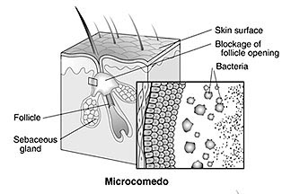
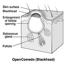
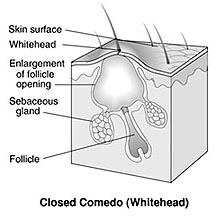
The three types of noninflammatory lesions caused by the accumulation of P. acnes in plugged follicles. From NIAMS |
Acne vulgaris is the most common disease caused by P. acnes. The four pathophysiologic features of acne, which occurs most often in the face, neck and back areas, are hyperkeratiniation, sebum production, bacterial proliferation, and inflammation. Three categories of lesions formed during acne are noninflammatory lesions, inflammatory lesions, and scars. Three noninflammatory lesions include microcomedos, open comedos, which have a central impaction of keratin and lipid and might or might not be slightly raised, and closed comedos, which are slightly elevated papules that could possibly form into a larger inflammatory lesion. Inflammatory lesions include more aggravated lesions such as pustules and large, fluctuant nodules.
Propionibacteria have been known to cause, although rarely, brain abscesses, subdural empyema, dental infections, endocarditis, continuous ambulatory peritoneal dialysis, conjunctivitis associated with contact lenses, and peritonitits (list from eMedicine). P. acnes is also associated with several types of infections and health problems such as anaerobic arthritis associated with prosthetic joints, and cases of osteomyelitits (rarely). P. acnes is also associated with certain spondyloarthropathies associated with florid acne vulgaris (isolated from bone foci and joints), and has been isolated as a cause of endocarditis (rarely) and reported as a cause of infectious keratitis when the cornea is compromised that can lead to vision loss.
Propionibacterium species can generally be treated with antibiotics used for the treatment of anaerobic infections; such antibiotics include penicillins, carbapenems, and clindamycin. However, recently there have been reports of antibiotic resistance in P. acnes.
Phages
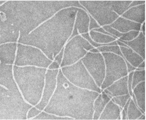
Until recently only Ff-related phages were known to be active in Gram-positive bacteria; however, at least five phages with filamentous morphology were shown to be active in P. freudenreichii. Filamentous phages, which are rod-shaped and contain circular single-stranded DNA (about 10 genes), generally infect Gram-negative bacteria using virion morphogenesis in which particles that do not cause lysis or cell death are secreted across the bacterial membranes continually. The five structural proteins of a filamentous phage are as follows: the major coat protein (pVIII) that forms the tube on the outer layer of the phage, two proteins (pVII and pIX) used for efficient particle assembly, and two proteins (pVI and pIII) used for particle stability and the ability of the phage to infect bacteria. The last four proteins (pVII, pIX, pVI, and pIII) are located at the ends of the rod-shaped phage. Two other proteins generally found in this type of phage (pI and pIV) are required for phage assembly and export. One of the filamentous phages that has been found to infect P. freudenreichii contains a ssDNA of 5,806 bases and has a G-C content of 64%, which is similar to the G-C content of P. freudenreichii. This phage does not contain a protein similar to pIV, which specifically assists in the outer membrane phage-conducting channel. This is probably due to the fact that since this phage infects Gram-positive bacteria, which have no outer membranes, the protein is not needed. (Chopin, et al. 2002)
References
Handa, Sajeev. "Propionibacterium Infections." 2002. eMedicine
