Uploads by Brlim
From MicrobeWiki, the student-edited microbiology resource
This special page shows all uploaded files.
| Date | Name | Thumbnail | Size | Description | Versions |
|---|---|---|---|---|---|
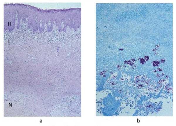
| 00:30, 27 August 2009 | 02-0485 1b.jpg (file) |  |
58 KB | a) Hematoxylin and eosin stain of a lesion specimen showing definitive Buruli ulcer disease in the preulcerative stage (original magnification 50x). Notice the psoriasiform epidermal hyperplasia (H), superficial dermal lichenoid inflammatory infiltrate (I | 1 |
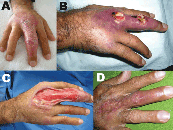
| 00:22, 27 August 2009 | 07-0904 2b.gif (file) |  |
128 KB | A) Nonulcerative edematous lesion on the right middle finger as first seen; B) ulcerated lesions on the right middle finger ≈4 weeks later; C) extensive debridement, 5.5 weeks after first seen; D) cured lesion 5 months after first seen, 1 month after au | 1 |
| 21:29, 26 August 2009 | Picture4004.jpg (file) |  |
24 KB | 1 | |
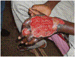
| 21:04, 26 August 2009 | Picture3004.jpg (file) |  |
24 KB | buruli ulcer on foot | 2 |
| 21:01, 26 August 2009 | Picture2004.gif (file) |  |
8 KB | owned | 1 |
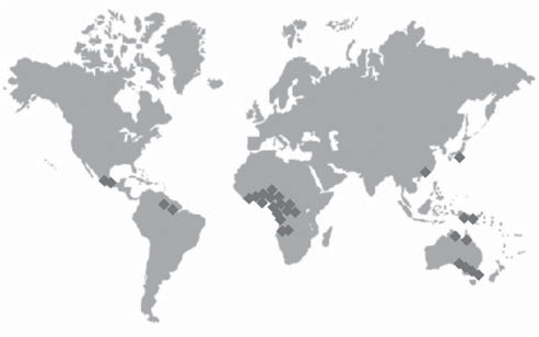
| 20:37, 26 August 2009 | Picture1004.png (file) |  |
35 KB | Regions affected by Buruli Ulcer | 1 |
