Uploads by Eldahank
From MicrobeWiki, the student-edited microbiology resource
This special page shows all uploaded files.
| Date | Name | Thumbnail | Size | Description | Versions |
|---|---|---|---|---|---|
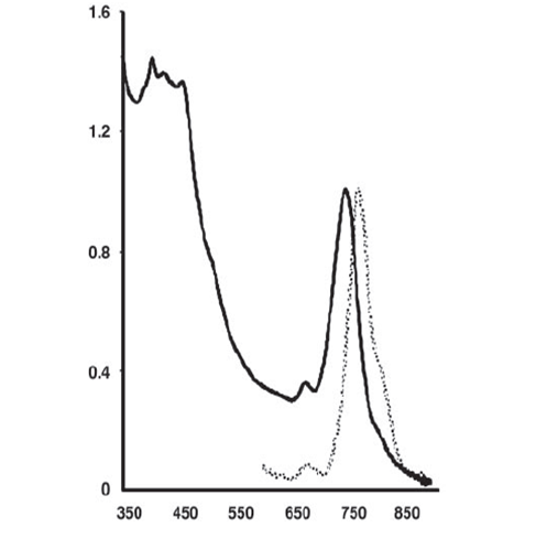
| 05:03, 1 May 2009 | Spectra data for GSB 1 Cells.png (file) |  |
51 KB | Figure 5. Absorption (solid line) and fluorescence emission (broken line) spectra of GSB1 intact cells. Vertical axis gives absorbance_fluorescence (arbitrary units) and horizontal axis gives wavelengths in nanometers. Image courtesy of Beatty et al. (3). | 1 |
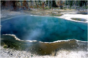
| 04:28, 16 April 2009 | Octopus Spring 1.png (file) |  |
165 KB | Octopus Spring. Image courtesy of http://serc.carleton.edu/microbelife/topics/octopusspring/index.html | 1 |
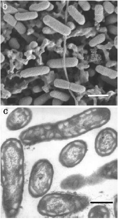
| 04:06, 16 April 2009 | New Sulfur Bacteria Microscopy 1.png (file) |  |
88 KB | Scanning Electron Microscopy of filter-deposited cells (B) and thin section Transmission Electron Microscopy (C). Note the electron transparent structures near the perimeter of the cell. These structures are characteristic of of chlorosomes. Bar=300nm.Ima | 1 |
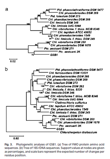
| 04:00, 16 April 2009 | New Sulfur Bacteria Phylogentic Tree 1.png (file) |  |
103 KB | Figure courtesy of Beatty, Blankenship, et al. (2005). | 1 |
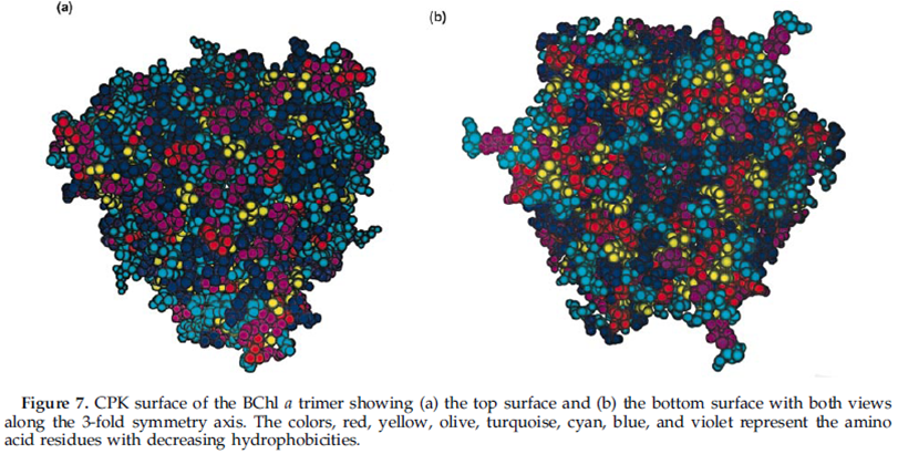
| 03:03, 16 April 2009 | Hydrophobicity of Amino Acids in BChl a 1.png (file) |  |
488 KB | Image courtesy of Zhou, Blankenship et al. (1997). | 2 |
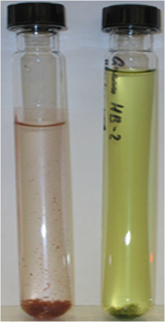
| 06:16, 15 April 2009 | Purple Sulfur vs. Green Sulfur Bacteria 1.png (file) |  |
398 KB | Tubes of purple (left) and green (right) sulfur bacteria. Image courtesy of the Yale University Department of Geology and Biophysics (http://earth.geology.yale.edu/~reb29/index.cgi?page-selection=5) | 1 |
| 05:55, 15 April 2009 | Light Harvesting Model of Green Sulfur Bacteria 1.png (file) |  |
468 KB | Model depicting the process of light harvesting in C. tepidum. The model includes the chlorosome, FMO protein, and reaction center. Photo courtesy of the University of Copenhagen Biology Department (http://www.bio.ku.dk/nuf/images/Ctepmodel040107_mod.jpg) | 1 |
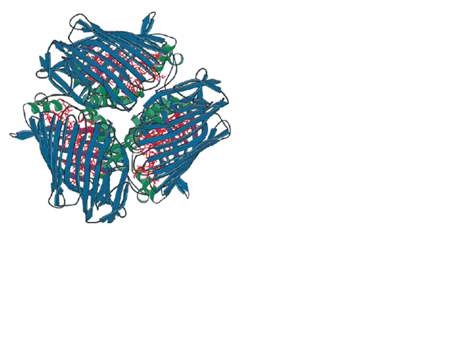
| 05:16, 15 April 2009 | BChl a Crystal Structure 2.png (file) |  |
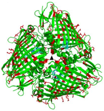
149 KB | BChl a Crystal Structure 1.png|frame|right|Ribbon diagram of the FMO (BChl a) protein from C. tepidum. Note the 3-fold symmetry of the three identical subunits. Image courtesy of the work conducted by Li, Zhou, Blankenship et al. (1997). | 1 |
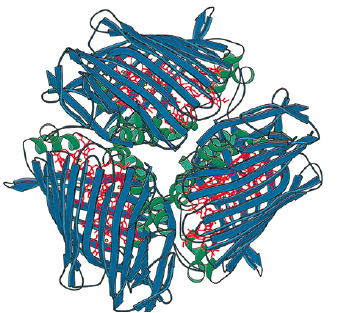
| 05:13, 15 April 2009 | BChl a Crystal Structure 1.png (file) |  |
330 KB | BChl a Crystal Structure 1.png|frame|right|Ribbon diagram of the FMO (BChl a) protein from C. tepidum. Note the 3-fold symmetry of the three identical subunits. Image courtesy of the work conducted by Li, Zhou, Blankenship et al. (1997). | 3 |
| 03:27, 14 April 2009 | FMO orientation taco.png (file) |  |
237 KB | Discovery of the orientation of the Fenna-Matthews-Olson protein in ''Chlorobium tepidum''. | 1 |
