Uploads by Map1934
From MicrobeWiki, the student-edited microbiology resource
This special page shows all uploaded files.
| Date | Name | Thumbnail | Size | Description | Versions |
|---|---|---|---|---|---|
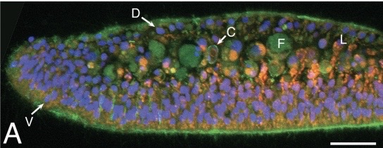
| 00:33, 13 December 2022 | 3AD2E51A-D488-41AD-AC2A-92ACC6052754 4 5005 c.jpeg (file) |  |
63 KB | This photo shows ''Trichoplax adhaerens'' as prepared by a microscope in a cross-section. The different stains allow different parts of the cell to be seen. In light green you see the autofluorescence. The bright green at the bottom shows filamentous actin that are present within epithelial cells. The blue-violet color depicts nuclei in epithelial cells. The reddish orange shows lipophil cells. Lastly, the pale green indicates the fiber cell bodies, more specifically their auto-fluorescent gr... | 1 |
