Uploads by Mhigashi
From MicrobeWiki, the student-edited microbiology resource
This special page shows all uploaded files.
| Date | Name | Thumbnail | Size | Description | Versions |
|---|---|---|---|---|---|
| 09:46, 28 August 2009 | 6511 lores.jpg (file) | 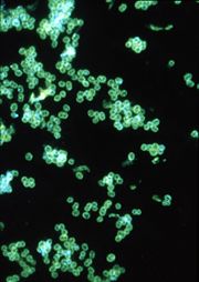 |
59 KB | This fluorescent antibody-stained micrograph depicts a positive result testing for the presence of gonorrhea. Courtesy of the CDC. | 1 |
| 08:07, 28 August 2009 | 10247 lores.jpg (file) | 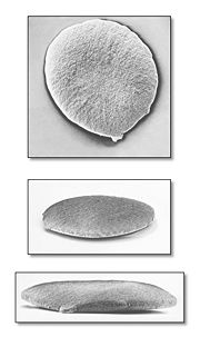 |
90 KB | This scanning electron micrograph (SEM) depicted three views of a single Gram-negative Neisseria gonorrhoeae bacterium. Under this highly-magnified view, the “roughened” texture of the bacterium’s cell wall is made visible. As a Gram-negative bacter | 1 |
| 07:26, 28 August 2009 | 5174 lores.jpg (file) | 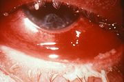 |
31 KB | This case of gonorrheal conjunctivitis resulted in partial blindness due to the spread of N. gonorrhoeae bacteria. Gonococci cause both localized infections, usually in the genital tract, and disseminated infections with seeding of various organs. Diagno | 1 |
| 07:10, 28 August 2009 | 651px-Condom rolled.jpg (file) | 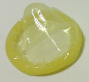 |
39 KB | If used properly, codom's provide 100% prevention against gonorrhea. | 1 |
| 06:31, 28 August 2009 | 401px-PatPong Dancer.jpg (file) |  |
64 KB | A dancer at a go-go bar along Patpong. Bangkok, Thailand. Courtesy of Wikipedia user Gregorof. Please credit Gregorof if used. | 1 |
| 06:16, 28 August 2009 | Neisseria gonorrhoeae PHIL 3693 lores.jpg (file) | 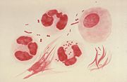 |
25 KB | This illustration depicts a urethral exudate containing Neisseria gonorrhoeae from a patient with gonococcal urethritis. This illustration depicts a Gram-stain of a urethral exudate showing typical intracellular gram-negative diplococci, and pleomorphic e | 1 |
| 05:53, 28 August 2009 | File-Damnoensaduak97.jpg (file) |  |
109 KB | Floating Market, Damnoen Saduak - Tahiland, 1997 | 1 |
