Uploads by Schwingel
From MicrobeWiki, the student-edited microbiology resource
Jump to navigationJump to search
This special page shows all uploaded files.
| Date | Name | Thumbnail | Size | Description | Versions |
|---|---|---|---|---|---|
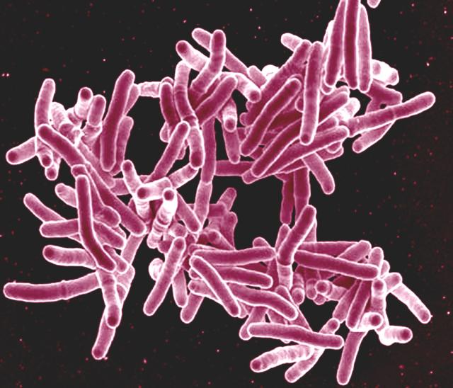
| 01:51, 15 April 2024 | M. tuberculosis.jpg (file) | Error creating thumbnail: File missing |
57 KB | Mycobacterium Tuberculosis Bacteria scanning electron micrograph Scanning electron micrograph of Mycobacterium tuberculosis bacteria. Credit: NIAID. Licensed under CC BY 2.0 DEED https://www.flickr.com/photos/niaid/5149398656 | 1 |
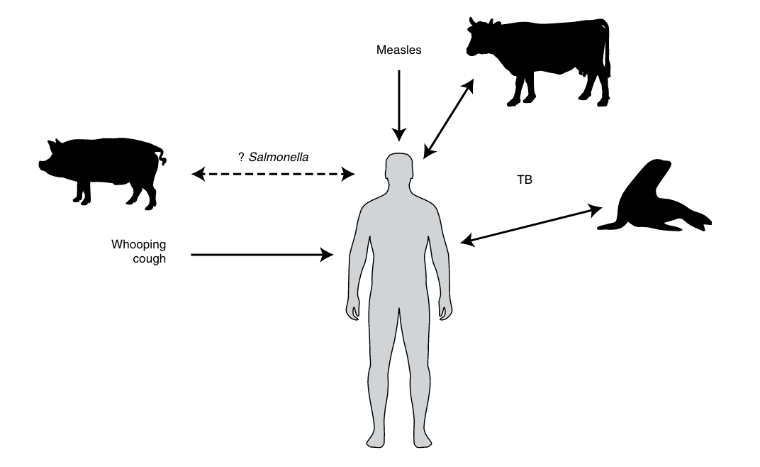
| 01:45, 15 April 2024 | Stone Domestic Origins Hypothesis.png (file) | Error creating thumbnail: Unable to save thumbnail to destination |
60 KB | The domestic origins for human disease hypothesis. This posits that agriculturalists living in close proximity with animals, especially livestock, would have been at high risk for zoonotic pathogens. From Stone, Anne C. 2020. “Getting Sick in the Neolithic.” Nature Ecology & Evolution 4 (3): 286–87. https://doi.org/10.1038/s41559-020-1115-8. | 1 |
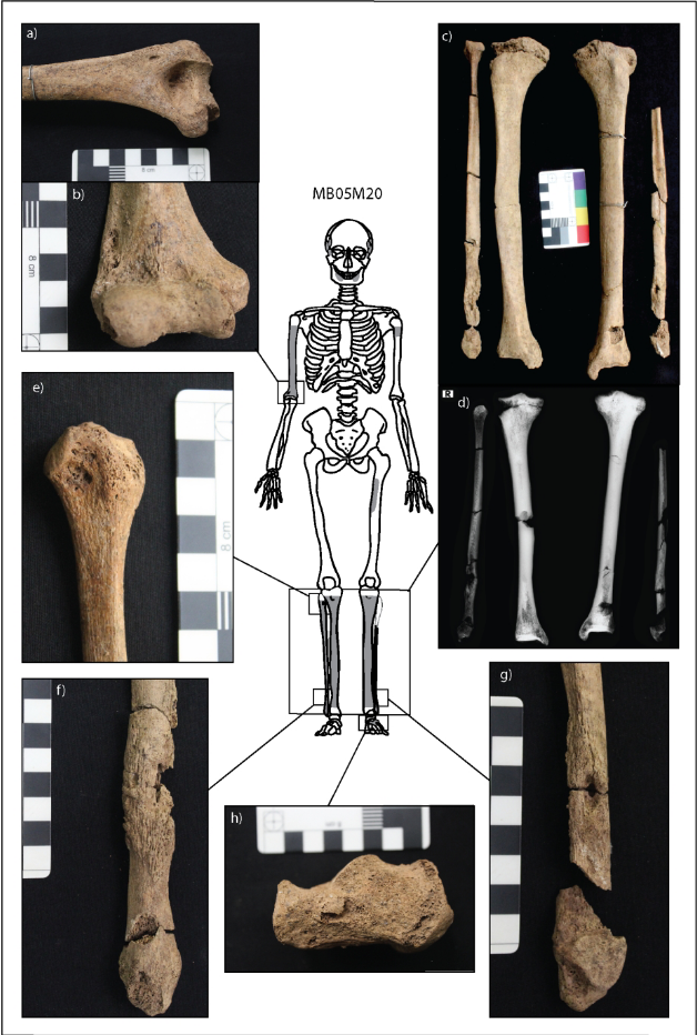
| 01:33, 15 April 2024 | Vlok et al. Fig 2 M20.png (file) | Error creating thumbnail: File missing |
788 KB | Lesions related to infectious disease in individual M20 from Neolithic Northern Vietnam. A) and b) show cavitations (holes) in the right humerus. C) and d) show new bone on the tibiae and fibulae. E), f), and g) show new bone growth and cavitations on the right fibula. H) shows cavitations on the left heel bone. From Vlok, Melandri, Marc Oxenham, Kate Domett, Tran Thi Minh, Thi Mai Huong Nguyen, Hirofumi Matsumura, Hiep Hoang Trinh, et al. 2020. “Two Probable Cases of Infection with Treponema... | 1 |
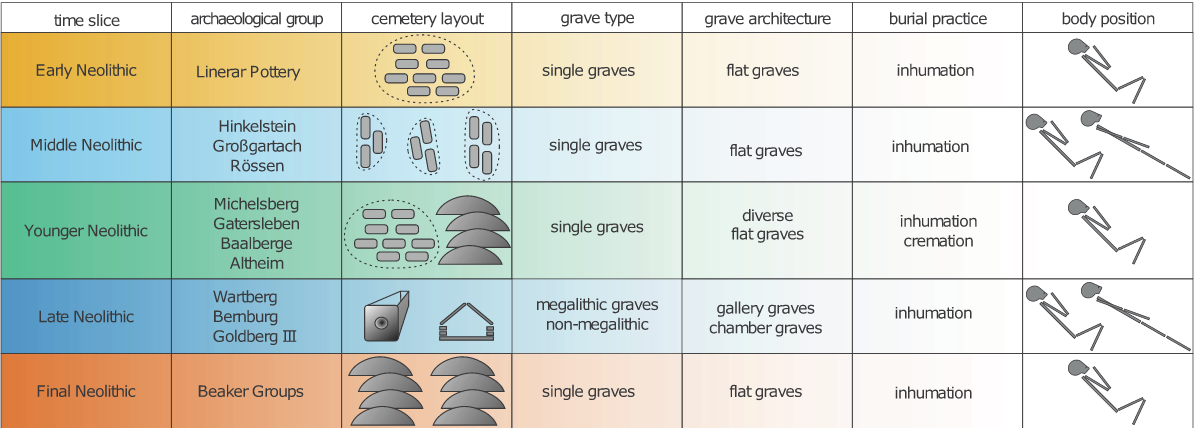
| 01:13, 15 April 2024 | Fuchs et al. Mortuary Practices Neolithic.png (file) | Error creating thumbnail: Unable to save thumbnail to destination |
214 KB | Fuchs et al. Figure 3: Archaeological groups, burials, and mortuary practices throughout the Neolithic in Germany. Image from Fuchs, Katharina, Christoph Rinne, Clara Drummer, Alexander Immel, Ben Krause-Kyora, and Almut Nebel. “Infectious Diseases and Neolithic Transformations: Evaluating Biological and Archaeological Proxies in the German Loess Zone between 5500 and 2500 BCE.” 2019. The Holocene 29 (10): 1545–57.http://journals.sagepub.com/doi/10.1177/0959683619857230 | 1 |
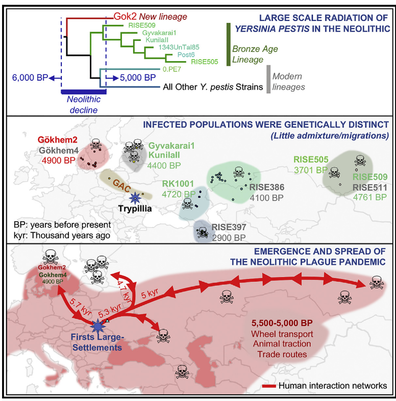
| 21:01, 14 April 2024 | Graphical Abstract Rascovan et al .png (file) | Error creating thumbnail: File missing |
661 KB | Graphical abstract describing the hypothesis of a Neolithic Yersinia pestis plague. Image from Rascovan, Nicolás, Karl-Göran Sjögren, Kristian Kristiansen, Rasmus Nielsen, Eske Willerslev, Christelle Desnues, and Simon Rasmussen. “Emergence and Spread of Basal Lineages of Yersinia Pestis during the Neolithic Decline.” 2019. Cell 176 (1–2): 295-305.e10. | 1 |
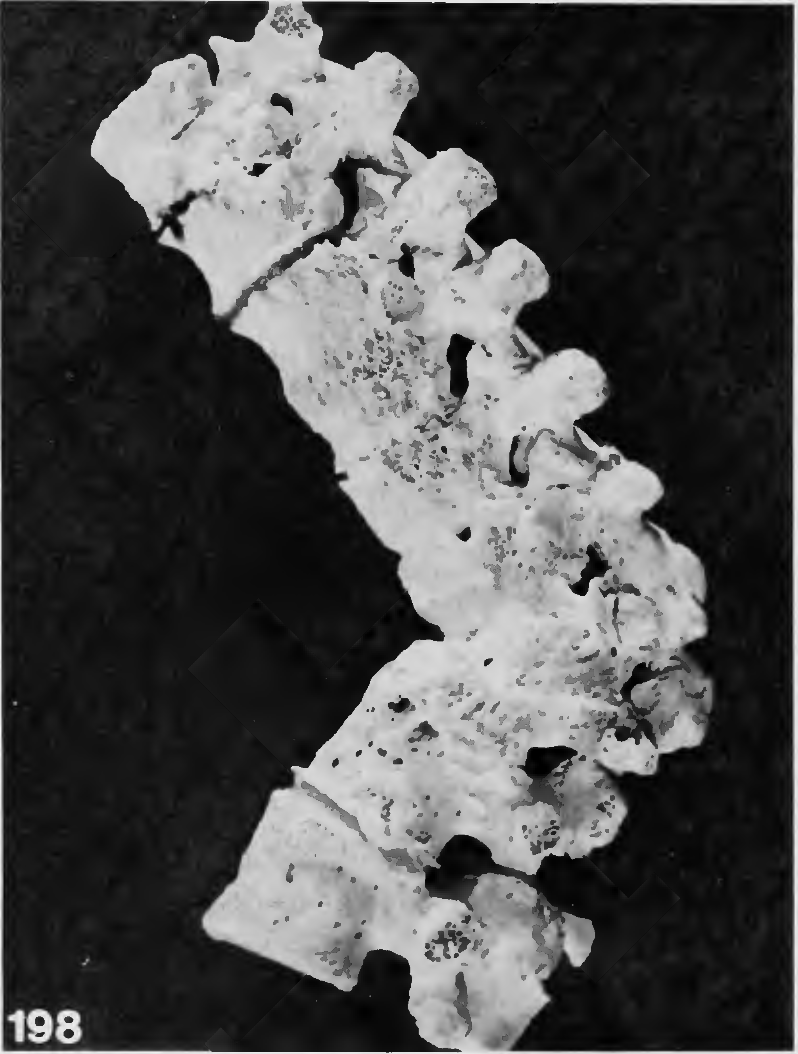
| 15:44, 14 April 2024 | Tuberculosis of Spine Buikstra.png (file) | Error creating thumbnail: File missing |
595 KB | Lateral view of spine affected by tuberculosis, partly healed, affecting thoracic vertebrae 7 and 8 and first lumbar vertebra. Image from Buikstra, J.E. ed. "Ortner's identification of pathological conditions in human skeletal remains." 2019. | 1 |
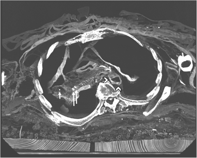
| 15:55, 23 February 2024 | Sabin et al Figure 1.png (file) | Error creating thumbnail: Unable to save thumbnail to destination |
179 KB | Figure 1 from Sabin et al. 2020. CT image of chest revealing calcifications suggesting Ranke complex and previous tuberculosis. Photo Credit:Genome Biology [https://genomebiology.biomedcentral.com/articles/10.1186/s13059-020-02112-1#Fig1]. | 1 |