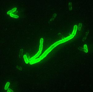
Yersinia pestis
A Microbial Biorealm page on the genus Yersinia pestis
Wiki
Classification
Higher order taxa
Kingdom: Eubacteria
Phylum: Proteobacteria
Class: Gamma Proteobacteria
Order: Enterobacteriale
Genus: Yersinia
Species
Yersinia pestis
Description and significance
Yersinia pestis was discovered in Hong Kong in 1894 by a Swiss physician Alexandre Yersin, who was a student of the Pasteur school of thought. He linked Y. pestis to the bubonic plauge, an epidemic that ravaged Europe during the 1300s. The organism was isolated during a outbreak in Hong Kong, a new geographical region for the organism that has been seen in Europe and Africa.
It is very important to have the genome sequenced for Y. pestis because this organism is capable of causing very fatal diseases. Since scientists were able to sequence the genome, they now have information of how diseases caused by this pathogenic bacteria develop and also the evolutionary history of the bacteria. Having the genome sequence also means that they are able to determine other species that are related to Yersinia pestis which can prevent future outbreaks.
Genome structure
Yersinia pestis has three subspecies and two have been sequenced, strain KIM and strain CO92. Each strain consists of one chromosome. Strain KIM consists of 4,600,755 base pairs and has a circular chromosome. "This strain is also related with the black plague. Strain CO92 has 4,653,728 base pairs and contains three plasmids (pMT1, pCD1 and pPCP1) of 96.2 kilobases (kb), 70.3 kb and 9.6 kb" (3). Strain KIM also carries these plasmids. These plasmids along with a pathogenicity island called HPI, create a protein that causes the pathogenicity of the organism. These factors are important for adhesion and injection of proteins into the host cell, invasion of the bacteria and binding of iron from red blood cells. The genome has many insertion sequences and many G-C base pair differences, which means frequent recombination. Many of the genes have been acquired from other bacteria and viruses. Strain CO92 also consists of 4,012 protein-coding genes and 150 pseudogenes (4).
A recent study compared both these strains to show that one of these strains may have evolved from the other throughout the years.
Cell structure and metabolism
Yersinia pestis is a rod shaped gram-negative bacteria that can also have a spherical shape. It is also covered by a slime envelope that is heat labile. When the bacteria is in a host, it is nonmotile (incapable of self-propelled movement), but when isolated it is motile (1).
Y. pestis uses aerobic respiration and anaerobic fermentation to produce and consume hydrogen gas for energy.
Ecology
Yersinia pestis interacts mainly with rodents such as rats and fleas. Through these carriers, Yersinia pestis is able to invade human cells and create diseases. Yersinia pestis are not rich in nuterients and can grow at temperatures ranging from about 26 Celcius to 37 Celcius.
Pathology
Y. pestis causes diseases through the bite of an infected rat or flea, but can also be transmitted by air. Fleas can become infected by taking the blood of other infected animals. Y. pestis grows in the midgut and eventually blocks the proventriculus, starving the flea for blood (1). The insects attempt to feed more often but end up giving back infected blood into the wound of the bite.
Symptons include:
- Sudden onset of high fever
- Emergence of a smooth, painful swelling of the lymph gland(s), called a buboe. The most common area is the groin, but swollen glands may also occur in the armpits or neck. Pain may occur in the area before the swelling.
- Chills
- General discomfort or ill feeling (malaise)
- Muscular pain
- Severe headache
- Seizures
"The major defense against Y pestis infection is the development of specific anti-envelope (F1) antibodies, which serve as opsonins for the virulent organisms, allowing their rapid phagocytosis and destruction while still within the initial infectious locus. The immune mechanism against this disease is extremely complex and involves a combination of humoral and cellular factors. The host is immune to virulent rechallenge, the inoculum being eliminated as though the organisms were completely avirulent". (1) Killed Y. pestis vaccines induce some measure of host protection, although it is less effective.
Yersinia pestis infections must be diagnosed quickly due to the high virulence of these organisms. Death from pneumonic plague can occur in as little as 24 hours after the first appearance of symptoms.
Yersinia pestis can be killed with mild heat(55°C) and by treatment with 0.5 percent phenol for 15 minutes. It is susceptible to sulfadiazine, streptomycin, tetracycline, and chloramphenicol.
Application to Biotechnology
This organism does not provide any enzymes or proteins for biotechnology.
Current Research
Researchers recently compared the strains of Yersinia pestis (strain C092 and KIM) and Yersinia pseudotuberculosis. This comparison would show how the pathogen could have evolved from each other within a few hundred years by acquiring new genes. These new genes can also inactivate some of the existing ones. In the end, Jargid et al determined that both strains could have diverged from a common ancestor thousands of years ago, however the lack of a reliable molecular clock, IS transposition and gene inactivation made it difficult to specifically determine the actual distance between both species (6). They also concluded that gene inactivation could have lead to the different genes.
Another research showed a recent emergence of new variants of Yersinia pestis in Madagascar. The researchers reported that the Y. pestis strain in Madagascar before 1982 was of the ribotype B, but after 1982 strains with ribotypes R, Q and T were isolated on a high plateau on the island (5).The researchers concluded that none of the strains studied from anywhere else in the world displayed these new ribotypes, that the new variants were not isolated on the seaport but on the high plateau and that the new strains were discovered after a recent plague surveillance was established.
Researchers have also detected the Yersinia pestis organism on a 400 year old set of teeth. They performed this research because the lack of suitable infected material has prevented direct discovery of the plague, thus making the idea of a black plague hypothetical. "The durability of dental pulp, together with its natural sterility, makes it a suitable material on which to conduct research" (2). They used bodies of people who died from the plague in Europe. "PCRs incorporating ancient DNA extracts and primers specific for the human beta-globin gene demonstrated the absence of inhibitors in these preparations. The incorporation of primers specific for Y. pestis rpoB and the recognized virulence-associated pla repeatedly yielded products that had a nucleotide sequence indistinguishable from that of modern day isolates of the bacterium" (2). The researchers were able to confirm that the disease was present at the end of the 16th century due to a nucleic acid-base confirmation. Many gram-negative bacteria (including E. tarda) use ferric uptake regulator (Fur) as an important transcriptional regulator. In E. tarda, Fur’s role is significant and multifaceted; Fur affects the growth, siderophore production, and acid tolerance of E. tarda, helps protect against oxidative stress and host serum, helps inhibit host immune response, and generally increases the overall virulence of the bacterium. Y. pestis is well known as a flea-borne bacteria. However, it evolved from Y. pseudotuberculosis, which exhibited significant oral toxicity to flea vectors. This oral toxicity is caused by urease activity which has been silenced in Y. pestis, due to expression of the mutant ureD allele. Restoration of functional ureD rendered Y. pestis toxic to vectors, which may be invaluable knowledge for decreasing plague transmission.
References
1. Collins, F.M., (1996). Pasteurella, Yersinia, and Francisella. In: Baron's Medical Microbiology (Baron S et al, eds.), 4th ed., Univ of Texas Medical Branch. ISBN 0-9631172-1-1.
2. Drancourt, M., et al.. (1998)."Detection of 400-year-old Yersinia pestis DNA in human dental pulp: An approach to the diagnosis of ancient septicemia". PNAS. 95 (21): 12637–12640. PMID 9770538.
3. Parkhill, J. et al.. (2001)."Genome sequence of Yersinia pestis, the causative agent of plague". Nature. 413: 523–527. PMID 11586360
4. Deng, W. et al.. (2002). "Genome Sequence of Yersinia pestis KIM". Journal of Bacteriology. 184 (16): 4601–4611. PMID 12142430.
5. Guiyoule, A. et al.. (1997) "Recent emergence of new variants of Yersinia pestis in Madagascar." Journal of Clinical Microbiology. 35 (11): 2826–2833.
6. Jangid, K. et al.. (2005) "Is Yersinia pestis a distinct species?" Current Science. 88 (5)
7. Center for Disease Controls Direct Fluorescent Antibody Stain (DFA), 2000x Magnification. CDC 2002 - US Government public domain image.
Edited by Danny Dang

