Yellow Fever Vaccine: Difference between revisions
No edit summary |
No edit summary |
||
| Line 5: | Line 5: | ||
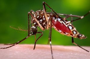
<br>Yellow fever is a human disease caused by the yellow fever virus, prevalent mostly in tropical climates. The yellow fever virus is a part of a family of viruses known as the Flavivirdae and the genus Flavivirus that fall under a broader category of arboviruses, or “arthropod-borne” viruses.<sup>1</sup> In this case, most common arthropod vector of YF is the <i>Aedes aegypti </i> mosquito. | |||
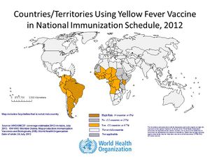
As stated previously, the disease is usually only prevalent in tropical regions of the world, mostly South America and parts of Africa (see figure 2). Even with vaccines available, yellow fever (YF) still causes 30,000 deaths annually with almost 200,000 cases occurring in Africa.<sup>2</sup> Moreover, YF is an acute infection, with mortality rates ranging from 25~50%.<sup>3</sup> Even worse, the number of cases of YF has increased in the past 20 years as a result of declining immunity among susceptible populations to infection, climate change, and urbanization. <sup>4</sup> Because of this, YF is a critical public health issue in South America and Africa. | |||
[[Image:YF_WHO_Map.jpg|thumb|300px|right|Figure 2. Map depicting geographic high risk areas for Yellow Fever, published by the WHO, July 2013.]] | |||
[[Image:YF_WHO_Map.jpg|thumb|300px|right|Figure 2. Map depicting geographic high-risk areas for Yellow Fever, published by the WHO, July 2013.]] | |||
==History== | ==History== | ||
<br> Spaniards documented the first major occurrence of YF in Yucatan, Mexico in 1648. <sup>5</sup> Local Mayans at the time referred to the disease as xekik (black-vomit), which was a common symptom of late stage YF. <sup>6</sup> Previous to arriving in Mexico however, it is hypothesized that the disease originated in Africa and spread as a result of slave ships travelling from Africa and transporting with them infected specimens of A. aegypti in their drinking water. <sup>7</sup> | |||
After the Yucatan outbreak, YF spread even further around the world causing high death tolls even in Barcelona and in the Mississippi Valley. <sup>8</sup> While it is now seen as a foreign tropical disease, there were numerous outbreaks in North America up through most of the 19th century, with epidemics in New York, 4,000 deaths in New Orleans, and even at one point an outbreak in Quebec. <sup>9</sup> | |||
It was not until 1881 when a Cuban scientist, Dr. Carlos Finlay, proposed that YF spread through A. aegypti that people began exterminating the mosquito and subsequently almost completely eradicated YF outside of South America and Africa. <sup>10</sup> The control of the disease was best exemplified by Cuba’s Major William C. Gorgas led and implemented specific mosquito-eradication procedures which effectively eradicated YF in the area within the year in 1901. <sup>11</sup> It was thanks to extermination efforts like Gorgas’ that Panama Canal could even be built considering many workers would have otherwise succumbed to YF as they worked on the canal. | |||
In 1937, Max Theiler created a live-attenuated vaccine for yellow fever by serial-passaging the YF virus through chicken embryotic tissue. <sup>12</sup> This vaccine, known as the 17D vaccine has impressively been highly effective in preventing the disease, with an unusually long antibody persistence upwards of 35 years in some individuals. <sup>13</sup> Since its production, over 500 million individuals have been vaccinated with the 17D vaccine further controlling the spread of yellow fever. <sup>14</sup> Appropriately, Max Thelier was rewarded with a Nobel Prize in 1951 for his contributions. | |||
| Line 18: | Line 28: | ||
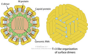
<br> The yellow fever virus (YFV) is a single, positive strand RNA virus with an icosahedral envelope as is characteristic of Flaviviruses and has a diameter of around 50nm. It has a genome of around 10kb. It is further characterized by the mature virions having three major types of structural proteins Capsid (C), Envelope (E), and Membrane (M), which make up the virion envelope. <sup>15</sup> Premature virions on the other hand have M precursor proteins (prM) that form heterodimers with E proteins, making the surface appear spiky.<sup>16</sup> Mature cells maintain a smooth surface as a result of the E proteins forming homodimers in “herringbone-like arrangements.” <sup>17</sup> | |||
<br> <u> Replication </u> | <br> <u> Replication </u> | ||
[[Image:Flavivirus_Life_Cycle.jpg|thumb|300px|right|Figure 4. Diagram of the Flavivirus' life cycle. Notice the initial binding to the host cell and the clathrin mediated endocytosis that follows the initial binding. Then the viral membrane fuses with the host's endosome to expose the viral ssRNA to the endoplasmic reticulum (ER). There, the ssRNA is transcribed and copied to make new viral proteins and new ssRNA. The viral particles are assembled and form a immature virion by budding off of the ER. After budding off, the immature virion moves to the Golgi. Moving from the Golgi to the host's membrane, the cell undergoes cleavages that cleave the prM protein into M proteins, maturing the virion before being secreted out of the host cell. Additionally, notice how the immature virion has a rough, spiked surface in contrast to the smooth surface of the mature virion. Diagram by Ted C. Pierson, NIH National Institute of Allergy and Infectious Diseases]] | [[Image:Flavivirus_Life_Cycle.jpg|thumb|300px|right|Figure 4. Diagram of the Flavivirus' life cycle. Notice the initial binding to the host cell and the clathrin mediated endocytosis that follows the initial binding. Then the viral membrane fuses with the host's endosome to expose the viral ssRNA to the endoplasmic reticulum (ER). There, the ssRNA is transcribed and copied to make new viral proteins and new ssRNA. The viral particles are assembled and form a immature virion by budding off of the ER. After budding off, the immature virion moves to the Golgi. Moving from the Golgi to the host's membrane, the cell undergoes cleavages that cleave the prM protein into M proteins, maturing the virion before being secreted out of the host cell. Additionally, notice how the immature virion has a rough, spiked surface in contrast to the smooth surface of the mature virion. Diagram by Ted C. Pierson, NIH National Institute of Allergy and Infectious Diseases]] | ||
<br>To replicate, the yellow fever virus first uses its E proteins to bind to receptors on the host cell to promote clatharin-mediated endocytosis. Then once the virion reaches the endosome in the eukaryotic cell, the YFV fuses its membrane with the host endosomal membrane and releases the RNA genome into the host’s cytoplasm. Once the single-stranded viral RNA (ssRNA) is released, it is translated and the translated proteins are brought to the endoplasmic reticulum (ER) to be assembled into immature virions. Specifically, the proteins are assembled in certain invaginations in the membrane of the ER called spherules. <sup>18</sup> At these spherules, the ssRNA is also replicated to create new positive sense ssRNA genomes for the new virions. | |||
Once the premature virions are formed, budding off of the ER, they move down the trans-Glogi network and are proteolytically cleaved, breaking down the prM and E heterodimers into M and E proteins to form mature virions. <sup>19</sup> Once mature, the virion particles leave the host cells through secretion. | |||
A large portion of the virion’s ability to replicate relies on the E proteins on its cell surface, which the virion depends on to bind to the host cell’s receptor proteins to enter the host cell as well as to induce fusion of the viral membrane with the host cell. Thus, a significant aspect of neutralizing the virus by the host’s immune system is to produce antibodies that target and disrupt the E protein’s ability to function. <sup>20</sup> | |||
| Line 29: | Line 43: | ||
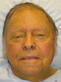
[[Image:Jaundice.jpg|thumb|200px|right|Figure 5. Example of a patient suffering from jaundice. Notice the distinct yellow pigmentation of the skin. Retrieved from wikimedia. Author:James Heilman, MD]] | [[Image:Jaundice.jpg|thumb|200px|right|Figure 5. Example of a patient suffering from jaundice. Notice the distinct yellow pigmentation of the skin. Retrieved from wikimedia. Author:James Heilman, MD]] | ||
<br>Yellow fever owes its name to its historically common symptom of causing jaundice in those who are afflicted. Jaundice is characterized by a yellow hue in an individual’s skin and eyes, as a result of excess bilirubin in the blood as the disease targets the liver. | |||
Female mosquitos carry the disease and individuals are only infected through their bite. For a mosquito to be able to pass on the disease however, it must first feed off of already infected individuals. Only after reaching a certain number of the yellow fever virus in its body, will the mosquito have their salivary glands infected and then begin transmitting the disease to its future blood feed hosts. | |||
The incubation period for disease is short and usually within 3~6 days after being bitten. <sup>21</sup> For most individuals, the extent of the illness is having a fever and flu-like symptoms such as nausea, back pain, and loss of appetite for upwards of a week. After these initial symptoms, the disease often passes and the survivors make a complete recovery and additionally develop a life-long immunity after infection. <sup>22</sup> | |||
However, for others, around 15% of those infected, the illness can enter a far more severe, secondary phase after the initial symptoms. <sup>23</sup> Once entering this stage, there is a greater than 20% likelihood of death. It is at this point in which symptoms such as jaundice (referred to earlier), bleeding, and organ failure. | |||
==Treatment and Cure== | ==Treatment and Cure== | ||
<br> Unfortunately, there are no treatments yet to cure yellow fever. Afflicted individuals are advised to be hospitalized and simply rest as much as possible. Some suggest using pain relievers to reduce the fever but outside of that, few medications are recommended. Specifically, anti-inflammatory drugs such as aspirin are to be avoided as they may worsen the symptoms and further promote bleeding. | |||
Besides care, health organizations strongly advise keeping the infected individuals away from any more contact with mosquitos. Since mosquitos must receive the yellow fever virus from feeding off of infected individuals to be contagious, by keeping an infected patient from being bitten by other mosquitos, it reduces the spread of the disease. | |||
==Mechanism of Infection== | ==Mechanism of Infection== | ||
<br> From examining tissue samples from those who died from the disease, studies have shown that the yellow fever virus appears to target hepatic cells, causing steatosis, apoptosis and necrosis in the hepatocytes. <sup>24</sup> More specifically, the virus tends to target hepatocytes in the midzonal area of the liver. <sup>25</sup> This targeting of a specific region of the liver has remained a puzzle for many years although studies are on going to better understand the cause of this targeting. Researchers are hopeful that by understanding what the virus are targeting, advancements can be made in treating patients already infected by the disease. | |||
An interesting observation that has been observed in studying the midzonal hepatocytes is that the much of the cell death in the area are attributed to apoptosis rather than lytic necrosis of the tissue. <sup>26</sup> Furthermore in the same study, although significant lesions were observed in the hepatocytes, analysis of the tissue showed very mild levels of inflammation. This disparity between the degree of hepatocyte injury and little to lack of inflammatory response is highly irregular. <sup>27</sup> Thus, many aspects of the actual pathological mechanism of the yellow fever virus remain unknown. | |||
==Vaccine== | |||
<br>A vaccine to prevent yellow fever has been developed and in use since the 1930s. Developed by Max Theiler, the chicken-embryo derived 17D vaccine has been a great success since it was first made available. However, in the more than 70 years of its existence, scientists have had very little understanding of what exactly has made the YF17D vaccine so effective, especially when an alternative vaccine was created around the same time by French scientists (the French neurotropic vaccine [FNV]) that failed to be as effective as the 17D vaccine. | |||
A recent study by Pulendran et al. (2006) sought to investigate the exact mechanism of the vaccine and its effectiveness. | |||
<br> <u>The Mechanism of Dendritic Cells (DC) and Immune Response</u> | |||
<br>A major component of the immune system and its response are based on the function of dendritic cells (DC) in sensing pathogenic threats. DCs are able to recognize pathogens and microbes through Toll-like receptors (TLRs). TLRs are a type of pathogen recognition receptor (PRR) and are well known for being highly conserved throughout many species. Specifically, in mammals, there are known to be at least 11 TLRs. Each of these TLRs recognizes certain components that are found in microbes, ranging from methylated DNA (TLR9) and lipopolysaccharides (TLR4). | |||
In terms of function, when a certain stimuli is recognized by a TLR, immature DCs are induced to maturate near the location of the stimuli and in maturing, move away to a T cell-rich area of the body. In these T cell rich areas, such as lymph nodes, the DCs stimulate T cells specific to the antigen that was initially recognized by the TLR. Thus, the TLRs and DCs together are crucial in activating the T cells and developing an individual’s immune memory. | |||
<br> <u>YF-17D and Immune Response</u> | |||
<br> When Pulendran et al. tested the activation of TLRs by the yellow fever 17D vaccine (YF-17D) on mice, they found that rather than stimulating just a single type of TLR, the vaccine in fact stimulated multiple TLRs. Specifically, they observed a significant activation of TLRs 2, 7, 8, and 9. Interestingly, while TLR2 and TLR7 were expected to activate (knowing TLR2 is activated by other viruses, and TLR7 is sensitive to single stranded RNA), TLR9 was unexpected, as it is known to only respond to unmethylated DNA. | |||
The researchers concluded that the major advantage and source of the effectiveness of the YF17D vaccine was its ability to activate multiple TLRs at once. They hypothesized that by having each of the TLRs 2, 7, 8, and 9 activate at once, the TLRs signaled their unique pathways together leading to a broader, more effective immune response. They also observed that one when even one of the TLRs was absent (in a mutant strain of mice) none of the TLRs was activated. One explanation of this is that the YF-17D vaccine in itself does not contain enough of any one type of molecule that would activate a TLR. However, by having some of many type of molecules and slightly stimulating multiple TLRs, the vaccine is able to induce a synergistic, full stimulation of multiple TLRs. | |||
<br> <u>T-Cell Balance</u> | |||
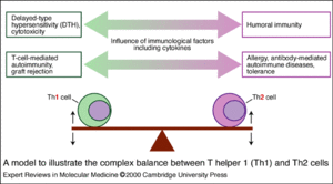
[[Image:Th1_and_Th2_Balance.gif|thumb|300px|right|Figure 6. Diagram depicting the balancing of Th1 and Th2 helper T-cells and their respective functions. Property of Cambridge University Press (2000)]] | [[Image:Th1_and_Th2_Balance.gif|thumb|300px|right|Figure 6. Diagram depicting the balancing of Th1 and Th2 helper T-cells and their respective functions. Property of Cambridge University Press (2000)]] | ||
<br> Another interesting facet of the study was the possible significance of the Th1/Th2 balance. Th1 and Th2 refer to two different forms of helper T-cells. Th1 helper cells promote inflammation and are ideal for fighting off viral infections. On the other hand, Th2 promote antibody production and more ideal for fighting off bacterial infections, toxins and allergens. | |||
In a perfectly healthy body, the two types of helper cells are usually balanced evenly and regulate the balance according to the needs of an individual. In Pulendran et al.’s study, they noticed certain mutations affected the balance of Th1/Th2 cells. Previous studies had shown that stimulation of most of the TLRs tended to lead to a significant Th1 response but stimulating TLR2 encouraged Th2 production slightly. Since TLRs 7, 8, and 9 as well TLR2 were observed to be stimulated, the study examined the Th1/Th2 after YF-17D vaccination. | |||
The studied observed that when the mutant strain of mice without MyD88, an “adaptor protein associated with all TLRs except TLR3 that mediates signal transduction,” <sup>28</sup> was given the YF-17D vaccine, rather than having an increased Th1 response, there was a significant inhibition of Th1. In another set of trials, when a mutant strain lacking TLR2 was vaccinated, the strain exhibited an enhanced Th1 response. Thus, it seems that TLR2 stimulation inhibits Th1 response but inhibits it in order to maintain a better Th1/Th2 balance. In other words, another contributing factor of the YF-17D vaccine’s effectiveness lies in stimulating the TLR2 in addition to the other TLRs to promote a beneficial balance of Th1 and Th2 helper T-cells. In fact, this balance may be the reason behind the long-lasting immunity that is characteristic of the YF-17D vaccine. | |||
<br> <u>“Clean” vs. “Dirty” Vaccines</u> | |||
<br> In line with the multiple TLRs stimulation conclusion, the study also proposed the possibility that a “dirty” vaccine is in fact more effective than a “clean” vaccine. To this day, the YF-17D vaccine is still produced in a very similar manner as when it was initially made, using chicken embryotic cell lines, and thus the vaccine contains chicken DNA, ovalbumin, and avian retroviral proteins in addition to YFV virions. <sup>29</sup> | |||
Having additional substances besides the actual vaccine cells may seem as a negative characteristic, making the vaccine “dirty.” But these additional particles in the vaccine may in fact be the reason why multiple TLRs are stimulated and consequently induce a better immune response in vaccinated individuals. This is very possible considering the vaccine containing viral ssRNA stimulated TLR9, which previously had only been know to be stimulated by unmethylated DNA. Thus, the study proposes that future developments of vaccines should focus on “having vaccine preparations that are ‘clean,’ but which recapitulate the immunogenicity of the ‘dirty’ vaccines.” <sup>30</sup> | |||
| Line 47: | Line 98: | ||
<br> <u>Chimera Vaccines</u> | <br> <u>Chimera Vaccines</u> | ||
<br> As a result of the effectiveness of the yellow fever vaccine 17D, a critical avenue of research has been to use the YF-17D cells and chimeric variants to produce vaccines against other arbovirus diseases such as dengue fever (DENV), West Nile (WNV), and Japanese encephalitis (JEV). Each of these three diseases presents an urgent public health issue. Currently, there have yet to be any vaccines developed for DENV or WNV further reinforcing the need for these vaccines. | |||
A new technology, called ChimeriVax has been developed and fine tuned in the last decade and has began to show promising results for making available a vaccine for these viruses. Specifically, with the ChimeriVax technology, scientists have been able to clone the YF-17D genome into cDNA and excise the specific genes that encode the E and prM proteins of the YFV and replace it with the gene for the E and prM protein of the target virus (ex. DENV). <sup>31</sup> Then, by transcribing the modified cDNA into RNA and growing the new virus in a cell culture, scientists have been able to create a new, chimeric virus that is designed to develop immunity against a different disease. Fortunately, promising chimeric vaccines for all three have been developed and await clinical trials before moving to commercial availability. | |||
<br> <u>Climate Change and Yellow Fever</u> | <br> <u>Climate Change and Yellow Fever</u> | ||
<br>Another current field of study is the effect of changing global climates on the susceptibility of the mosquitos (A. aegypti) to the yellow fever virus and consequently, the possibility of increasing transmission to humans. A greater portion of research into the yellow fever virus focuses on medical interests and how the virus interacts with humans. However, another portion of understanding YFV is the susceptibility of A. aegypti to the virus, considering if we can prevent the spread of the disease in mosquitos, it will be far easier to prevent it human transmission. With that in mind, some research has shown that to a certain degree, higher temperatures correlate with a higher infection rate of the virus. <sup>32</sup> A study showed that the extrinsic incubation period (EIP), or the time it takes between being infected by a blood meal and oral transmission, is inversely related to the temperature. Specifically, the study observed the EIP of the yellow fever virus in a certain species of mosquitos was 28 days when the temperature was 25 <sup>o</sup>C while only 12 days at 30 <sup>o</sup>C. <sup>33</sup> Therefore, if global climate change continues to progress at the current rate, it may mean a higher likelihood of yellow fever transmission as well as other arbovirus-based diseases. | |||
<br> <u>Availability of Yellow Fever 17D Vaccine</u> | <br> <u>Availability of Yellow Fever 17D Vaccine</u> | ||
<br> An increasing concern regarding the YF-17D vaccine is the availability of the vaccine worldwide. <sup>34</sup> Luckily, in recent years there have not been overwhelmingly numerous outbreaks of yellow fever around world. However, because the vaccine still relies on the same 70+ year old, out of date method to create the vaccine using chicken embryos, there is a very real concern that if multiple large-scale outbreaks were to occur at once, stockpiles of the vaccine would be exhausted and production could not keep up with the demand with short notice. Thus, the need for an improved method of creating the vaccine that is not only more efficient but also is able to keep the same effectiveness as the current YF-17D vaccine is necessary. | |||
==References== | |||
<sup>1, 5, 6, 9, 11, 34</sup> Barrett, A. D. T., & Higgs, S. (2007). Yellow fever: A disease that has yet to be conquered. Annual Review of Entomology, 52(1), 209-229. doi:10.1146/annurev.ento.52.110405.091454 | |||
<sup>3, 7, 8, 10</sup> Michael A. Tolle, Mosquito-borne Diseases, Current Problems in Pediatric and Adolescent Health Care, Volume 39, Issue 4, April 2009, Pages 97-140, ISSN 1538-5442, http://dx.doi.org/10.1016/j.cppeds.2009.01.001. (http://www.sciencedirect.com/science/article/pii/S1538544209000145) | |||
<sup>2</sup> Yellow fever. (n.d.). WHO. Retrieved April 21, 2014, from http://www.who.int/mediacentre/factsheets/fs100/en/ | |||
<sup>12, 14, 15, 16, 17, 19, 20</sup> Heinz, F. X., & Stiasny, K. (2012). Flaviviruses and flavivirus vaccines. Vaccine, 30(29), 4301-4306. doi:10.1016/j.vaccine.2011.09.114 (http://www.sciencedirect.com/science/article/pii/S0264410X11015568) | |||
<sup>18</sup> ViralZone: Flavivirus. (n.d.). Expasy: ViralZone. Retrieved April 21, 2014, from http://viralzone.expasy.org/all_by_species/24.html | |||
<sup>21, 22, 23</sup> Yellow Fever: Symptoms and Treatment. (2011, December 13). Centers for Disease Control and Prevention. Retrieved April 21, 2014, from http://www.cdc.gov/yellowfever/symptoms/index.html | |||
<sup>24, 25, 26, 27</sup> Quaresma, J. A. S., Barros, Vera L R S, Pagliari, C., Fernandes, E. R., Guedes, F., Takakura, C. F. H., Duarte, M. I. S. (2006). Revisiting the liver in human yellow fever: Virus-induced apoptosis in hepatocytes associated with TGF-β, TNF-α and NK cells activity. Virology,345(1), 22-30. doi:10.1016/j.virol.2005.09.058 | |||
<sup>32, 33</sup> William C. Black IV, Kristine E. Bennett, Norma Gorrochótegui-Escalante, Carolina V. Barillas-Mury, Ildefonso Fernández-Salas, Marı́a de Lourdes Muñoz, José A. Farfán-Alé, Ken E. Olson, Barry J. Beaty, Flavivirus Susceptibility in Aedes aegypti, Archives of Medical Research, Volume 33, Issue 4, July–August 2002, Pages 379-388, ISSN 0188-4409, http://dx.doi.org/10.1016/S0188-4409(02)00373-9. (http://www.sciencedirect.com/science/article/pii/S0188440902003739) | |||
<sup>13, 28, 29, 30</sup> Querec, T. et al. Yellow fever vaccine YF-17D activates multiple dendritic cell subsets via TLR2, 7, 8, and 9 to stimulate polyvalent immunity. J. Exp. Med. 203, 413–424 (2006) | |||
<sup>31</sup> Bruno Guy, Farshad Guirakhoo, Veronique Barban, Stephen Higgs, Thomas P. Monath, Jean Lang, Preclinical and clinical development of YFV 17D-based chimeric vaccines against dengue, West Nile and Japanese encephalitis viruses, Vaccine, Volume 28, Issue 3, 8 January 2010, Pages 632-649, ISSN 0264-410X, http://dx.doi.org/10.1016/j.vaccine.2009.09.098. (http://www.sciencedirect.com/science/article/pii/S0264410X09014455) | |||
Edited by Christopher Kei Helm of [mailto:slonczewski@kenyon.edu Joan Slonczewski] for [http://biology.kenyon.edu/courses/biol238/biol238syl14.html BIOL 238 Microbiology], 2014, [http://www.kenyon.edu/index.xml Kenyon College]. | |||
<!--Do not edit or remove this line-->[[Category:Pages edited by students of Joan Slonczewski at Kenyon College]] | |||
Revision as of 00:16, 23 April 2014
By Christopher Kei Helm
Overview
Yellow fever is a human disease caused by the yellow fever virus, prevalent mostly in tropical climates. The yellow fever virus is a part of a family of viruses known as the Flavivirdae and the genus Flavivirus that fall under a broader category of arboviruses, or “arthropod-borne” viruses.1 In this case, most common arthropod vector of YF is the Aedes aegypti mosquito.
As stated previously, the disease is usually only prevalent in tropical regions of the world, mostly South America and parts of Africa (see figure 2). Even with vaccines available, yellow fever (YF) still causes 30,000 deaths annually with almost 200,000 cases occurring in Africa.2 Moreover, YF is an acute infection, with mortality rates ranging from 25~50%.3 Even worse, the number of cases of YF has increased in the past 20 years as a result of declining immunity among susceptible populations to infection, climate change, and urbanization. 4 Because of this, YF is a critical public health issue in South America and Africa.
History
Spaniards documented the first major occurrence of YF in Yucatan, Mexico in 1648. 5 Local Mayans at the time referred to the disease as xekik (black-vomit), which was a common symptom of late stage YF. 6 Previous to arriving in Mexico however, it is hypothesized that the disease originated in Africa and spread as a result of slave ships travelling from Africa and transporting with them infected specimens of A. aegypti in their drinking water. 7
After the Yucatan outbreak, YF spread even further around the world causing high death tolls even in Barcelona and in the Mississippi Valley. 8 While it is now seen as a foreign tropical disease, there were numerous outbreaks in North America up through most of the 19th century, with epidemics in New York, 4,000 deaths in New Orleans, and even at one point an outbreak in Quebec. 9
It was not until 1881 when a Cuban scientist, Dr. Carlos Finlay, proposed that YF spread through A. aegypti that people began exterminating the mosquito and subsequently almost completely eradicated YF outside of South America and Africa. 10 The control of the disease was best exemplified by Cuba’s Major William C. Gorgas led and implemented specific mosquito-eradication procedures which effectively eradicated YF in the area within the year in 1901. 11 It was thanks to extermination efforts like Gorgas’ that Panama Canal could even be built considering many workers would have otherwise succumbed to YF as they worked on the canal.
In 1937, Max Theiler created a live-attenuated vaccine for yellow fever by serial-passaging the YF virus through chicken embryotic tissue. 12 This vaccine, known as the 17D vaccine has impressively been highly effective in preventing the disease, with an unusually long antibody persistence upwards of 35 years in some individuals. 13 Since its production, over 500 million individuals have been vaccinated with the 17D vaccine further controlling the spread of yellow fever. 14 Appropriately, Max Thelier was rewarded with a Nobel Prize in 1951 for his contributions.
Virus
The yellow fever virus (YFV) is a single, positive strand RNA virus with an icosahedral envelope as is characteristic of Flaviviruses and has a diameter of around 50nm. It has a genome of around 10kb. It is further characterized by the mature virions having three major types of structural proteins Capsid (C), Envelope (E), and Membrane (M), which make up the virion envelope. 15 Premature virions on the other hand have M precursor proteins (prM) that form heterodimers with E proteins, making the surface appear spiky.16 Mature cells maintain a smooth surface as a result of the E proteins forming homodimers in “herringbone-like arrangements.” 17
Replication
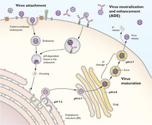
To replicate, the yellow fever virus first uses its E proteins to bind to receptors on the host cell to promote clatharin-mediated endocytosis. Then once the virion reaches the endosome in the eukaryotic cell, the YFV fuses its membrane with the host endosomal membrane and releases the RNA genome into the host’s cytoplasm. Once the single-stranded viral RNA (ssRNA) is released, it is translated and the translated proteins are brought to the endoplasmic reticulum (ER) to be assembled into immature virions. Specifically, the proteins are assembled in certain invaginations in the membrane of the ER called spherules. 18 At these spherules, the ssRNA is also replicated to create new positive sense ssRNA genomes for the new virions.
Once the premature virions are formed, budding off of the ER, they move down the trans-Glogi network and are proteolytically cleaved, breaking down the prM and E heterodimers into M and E proteins to form mature virions. 19 Once mature, the virion particles leave the host cells through secretion.
A large portion of the virion’s ability to replicate relies on the E proteins on its cell surface, which the virion depends on to bind to the host cell’s receptor proteins to enter the host cell as well as to induce fusion of the viral membrane with the host cell. Thus, a significant aspect of neutralizing the virus by the host’s immune system is to produce antibodies that target and disrupt the E protein’s ability to function. 20
Symptoms
Yellow fever owes its name to its historically common symptom of causing jaundice in those who are afflicted. Jaundice is characterized by a yellow hue in an individual’s skin and eyes, as a result of excess bilirubin in the blood as the disease targets the liver.
Female mosquitos carry the disease and individuals are only infected through their bite. For a mosquito to be able to pass on the disease however, it must first feed off of already infected individuals. Only after reaching a certain number of the yellow fever virus in its body, will the mosquito have their salivary glands infected and then begin transmitting the disease to its future blood feed hosts.
The incubation period for disease is short and usually within 3~6 days after being bitten. 21 For most individuals, the extent of the illness is having a fever and flu-like symptoms such as nausea, back pain, and loss of appetite for upwards of a week. After these initial symptoms, the disease often passes and the survivors make a complete recovery and additionally develop a life-long immunity after infection. 22
However, for others, around 15% of those infected, the illness can enter a far more severe, secondary phase after the initial symptoms. 23 Once entering this stage, there is a greater than 20% likelihood of death. It is at this point in which symptoms such as jaundice (referred to earlier), bleeding, and organ failure.
Treatment and Cure
Unfortunately, there are no treatments yet to cure yellow fever. Afflicted individuals are advised to be hospitalized and simply rest as much as possible. Some suggest using pain relievers to reduce the fever but outside of that, few medications are recommended. Specifically, anti-inflammatory drugs such as aspirin are to be avoided as they may worsen the symptoms and further promote bleeding.
Besides care, health organizations strongly advise keeping the infected individuals away from any more contact with mosquitos. Since mosquitos must receive the yellow fever virus from feeding off of infected individuals to be contagious, by keeping an infected patient from being bitten by other mosquitos, it reduces the spread of the disease.
Mechanism of Infection
From examining tissue samples from those who died from the disease, studies have shown that the yellow fever virus appears to target hepatic cells, causing steatosis, apoptosis and necrosis in the hepatocytes. 24 More specifically, the virus tends to target hepatocytes in the midzonal area of the liver. 25 This targeting of a specific region of the liver has remained a puzzle for many years although studies are on going to better understand the cause of this targeting. Researchers are hopeful that by understanding what the virus are targeting, advancements can be made in treating patients already infected by the disease.
An interesting observation that has been observed in studying the midzonal hepatocytes is that the much of the cell death in the area are attributed to apoptosis rather than lytic necrosis of the tissue. 26 Furthermore in the same study, although significant lesions were observed in the hepatocytes, analysis of the tissue showed very mild levels of inflammation. This disparity between the degree of hepatocyte injury and little to lack of inflammatory response is highly irregular. 27 Thus, many aspects of the actual pathological mechanism of the yellow fever virus remain unknown.
Vaccine
A vaccine to prevent yellow fever has been developed and in use since the 1930s. Developed by Max Theiler, the chicken-embryo derived 17D vaccine has been a great success since it was first made available. However, in the more than 70 years of its existence, scientists have had very little understanding of what exactly has made the YF17D vaccine so effective, especially when an alternative vaccine was created around the same time by French scientists (the French neurotropic vaccine [FNV]) that failed to be as effective as the 17D vaccine.
A recent study by Pulendran et al. (2006) sought to investigate the exact mechanism of the vaccine and its effectiveness.
The Mechanism of Dendritic Cells (DC) and Immune Response
A major component of the immune system and its response are based on the function of dendritic cells (DC) in sensing pathogenic threats. DCs are able to recognize pathogens and microbes through Toll-like receptors (TLRs). TLRs are a type of pathogen recognition receptor (PRR) and are well known for being highly conserved throughout many species. Specifically, in mammals, there are known to be at least 11 TLRs. Each of these TLRs recognizes certain components that are found in microbes, ranging from methylated DNA (TLR9) and lipopolysaccharides (TLR4).
In terms of function, when a certain stimuli is recognized by a TLR, immature DCs are induced to maturate near the location of the stimuli and in maturing, move away to a T cell-rich area of the body. In these T cell rich areas, such as lymph nodes, the DCs stimulate T cells specific to the antigen that was initially recognized by the TLR. Thus, the TLRs and DCs together are crucial in activating the T cells and developing an individual’s immune memory.
YF-17D and Immune Response
When Pulendran et al. tested the activation of TLRs by the yellow fever 17D vaccine (YF-17D) on mice, they found that rather than stimulating just a single type of TLR, the vaccine in fact stimulated multiple TLRs. Specifically, they observed a significant activation of TLRs 2, 7, 8, and 9. Interestingly, while TLR2 and TLR7 were expected to activate (knowing TLR2 is activated by other viruses, and TLR7 is sensitive to single stranded RNA), TLR9 was unexpected, as it is known to only respond to unmethylated DNA.
The researchers concluded that the major advantage and source of the effectiveness of the YF17D vaccine was its ability to activate multiple TLRs at once. They hypothesized that by having each of the TLRs 2, 7, 8, and 9 activate at once, the TLRs signaled their unique pathways together leading to a broader, more effective immune response. They also observed that one when even one of the TLRs was absent (in a mutant strain of mice) none of the TLRs was activated. One explanation of this is that the YF-17D vaccine in itself does not contain enough of any one type of molecule that would activate a TLR. However, by having some of many type of molecules and slightly stimulating multiple TLRs, the vaccine is able to induce a synergistic, full stimulation of multiple TLRs.
T-Cell Balance
Another interesting facet of the study was the possible significance of the Th1/Th2 balance. Th1 and Th2 refer to two different forms of helper T-cells. Th1 helper cells promote inflammation and are ideal for fighting off viral infections. On the other hand, Th2 promote antibody production and more ideal for fighting off bacterial infections, toxins and allergens.
In a perfectly healthy body, the two types of helper cells are usually balanced evenly and regulate the balance according to the needs of an individual. In Pulendran et al.’s study, they noticed certain mutations affected the balance of Th1/Th2 cells. Previous studies had shown that stimulation of most of the TLRs tended to lead to a significant Th1 response but stimulating TLR2 encouraged Th2 production slightly. Since TLRs 7, 8, and 9 as well TLR2 were observed to be stimulated, the study examined the Th1/Th2 after YF-17D vaccination.
The studied observed that when the mutant strain of mice without MyD88, an “adaptor protein associated with all TLRs except TLR3 that mediates signal transduction,” 28 was given the YF-17D vaccine, rather than having an increased Th1 response, there was a significant inhibition of Th1. In another set of trials, when a mutant strain lacking TLR2 was vaccinated, the strain exhibited an enhanced Th1 response. Thus, it seems that TLR2 stimulation inhibits Th1 response but inhibits it in order to maintain a better Th1/Th2 balance. In other words, another contributing factor of the YF-17D vaccine’s effectiveness lies in stimulating the TLR2 in addition to the other TLRs to promote a beneficial balance of Th1 and Th2 helper T-cells. In fact, this balance may be the reason behind the long-lasting immunity that is characteristic of the YF-17D vaccine.
“Clean” vs. “Dirty” Vaccines
In line with the multiple TLRs stimulation conclusion, the study also proposed the possibility that a “dirty” vaccine is in fact more effective than a “clean” vaccine. To this day, the YF-17D vaccine is still produced in a very similar manner as when it was initially made, using chicken embryotic cell lines, and thus the vaccine contains chicken DNA, ovalbumin, and avian retroviral proteins in addition to YFV virions. 29
Having additional substances besides the actual vaccine cells may seem as a negative characteristic, making the vaccine “dirty.” But these additional particles in the vaccine may in fact be the reason why multiple TLRs are stimulated and consequently induce a better immune response in vaccinated individuals. This is very possible considering the vaccine containing viral ssRNA stimulated TLR9, which previously had only been know to be stimulated by unmethylated DNA. Thus, the study proposes that future developments of vaccines should focus on “having vaccine preparations that are ‘clean,’ but which recapitulate the immunogenicity of the ‘dirty’ vaccines.” 30
Future Research
Chimera Vaccines
As a result of the effectiveness of the yellow fever vaccine 17D, a critical avenue of research has been to use the YF-17D cells and chimeric variants to produce vaccines against other arbovirus diseases such as dengue fever (DENV), West Nile (WNV), and Japanese encephalitis (JEV). Each of these three diseases presents an urgent public health issue. Currently, there have yet to be any vaccines developed for DENV or WNV further reinforcing the need for these vaccines.
A new technology, called ChimeriVax has been developed and fine tuned in the last decade and has began to show promising results for making available a vaccine for these viruses. Specifically, with the ChimeriVax technology, scientists have been able to clone the YF-17D genome into cDNA and excise the specific genes that encode the E and prM proteins of the YFV and replace it with the gene for the E and prM protein of the target virus (ex. DENV). 31 Then, by transcribing the modified cDNA into RNA and growing the new virus in a cell culture, scientists have been able to create a new, chimeric virus that is designed to develop immunity against a different disease. Fortunately, promising chimeric vaccines for all three have been developed and await clinical trials before moving to commercial availability.
Climate Change and Yellow Fever
Another current field of study is the effect of changing global climates on the susceptibility of the mosquitos (A. aegypti) to the yellow fever virus and consequently, the possibility of increasing transmission to humans. A greater portion of research into the yellow fever virus focuses on medical interests and how the virus interacts with humans. However, another portion of understanding YFV is the susceptibility of A. aegypti to the virus, considering if we can prevent the spread of the disease in mosquitos, it will be far easier to prevent it human transmission. With that in mind, some research has shown that to a certain degree, higher temperatures correlate with a higher infection rate of the virus. 32 A study showed that the extrinsic incubation period (EIP), or the time it takes between being infected by a blood meal and oral transmission, is inversely related to the temperature. Specifically, the study observed the EIP of the yellow fever virus in a certain species of mosquitos was 28 days when the temperature was 25 oC while only 12 days at 30 oC. 33 Therefore, if global climate change continues to progress at the current rate, it may mean a higher likelihood of yellow fever transmission as well as other arbovirus-based diseases.
Availability of Yellow Fever 17D Vaccine
An increasing concern regarding the YF-17D vaccine is the availability of the vaccine worldwide. 34 Luckily, in recent years there have not been overwhelmingly numerous outbreaks of yellow fever around world. However, because the vaccine still relies on the same 70+ year old, out of date method to create the vaccine using chicken embryos, there is a very real concern that if multiple large-scale outbreaks were to occur at once, stockpiles of the vaccine would be exhausted and production could not keep up with the demand with short notice. Thus, the need for an improved method of creating the vaccine that is not only more efficient but also is able to keep the same effectiveness as the current YF-17D vaccine is necessary.
References
1, 5, 6, 9, 11, 34 Barrett, A. D. T., & Higgs, S. (2007). Yellow fever: A disease that has yet to be conquered. Annual Review of Entomology, 52(1), 209-229. doi:10.1146/annurev.ento.52.110405.091454 3, 7, 8, 10 Michael A. Tolle, Mosquito-borne Diseases, Current Problems in Pediatric and Adolescent Health Care, Volume 39, Issue 4, April 2009, Pages 97-140, ISSN 1538-5442, http://dx.doi.org/10.1016/j.cppeds.2009.01.001. (http://www.sciencedirect.com/science/article/pii/S1538544209000145)
2 Yellow fever. (n.d.). WHO. Retrieved April 21, 2014, from http://www.who.int/mediacentre/factsheets/fs100/en/
12, 14, 15, 16, 17, 19, 20 Heinz, F. X., & Stiasny, K. (2012). Flaviviruses and flavivirus vaccines. Vaccine, 30(29), 4301-4306. doi:10.1016/j.vaccine.2011.09.114 (http://www.sciencedirect.com/science/article/pii/S0264410X11015568)
18 ViralZone: Flavivirus. (n.d.). Expasy: ViralZone. Retrieved April 21, 2014, from http://viralzone.expasy.org/all_by_species/24.html
21, 22, 23 Yellow Fever: Symptoms and Treatment. (2011, December 13). Centers for Disease Control and Prevention. Retrieved April 21, 2014, from http://www.cdc.gov/yellowfever/symptoms/index.html
24, 25, 26, 27 Quaresma, J. A. S., Barros, Vera L R S, Pagliari, C., Fernandes, E. R., Guedes, F., Takakura, C. F. H., Duarte, M. I. S. (2006). Revisiting the liver in human yellow fever: Virus-induced apoptosis in hepatocytes associated with TGF-β, TNF-α and NK cells activity. Virology,345(1), 22-30. doi:10.1016/j.virol.2005.09.058
32, 33 William C. Black IV, Kristine E. Bennett, Norma Gorrochótegui-Escalante, Carolina V. Barillas-Mury, Ildefonso Fernández-Salas, Marı́a de Lourdes Muñoz, José A. Farfán-Alé, Ken E. Olson, Barry J. Beaty, Flavivirus Susceptibility in Aedes aegypti, Archives of Medical Research, Volume 33, Issue 4, July–August 2002, Pages 379-388, ISSN 0188-4409, http://dx.doi.org/10.1016/S0188-4409(02)00373-9. (http://www.sciencedirect.com/science/article/pii/S0188440902003739)
13, 28, 29, 30 Querec, T. et al. Yellow fever vaccine YF-17D activates multiple dendritic cell subsets via TLR2, 7, 8, and 9 to stimulate polyvalent immunity. J. Exp. Med. 203, 413–424 (2006)
31 Bruno Guy, Farshad Guirakhoo, Veronique Barban, Stephen Higgs, Thomas P. Monath, Jean Lang, Preclinical and clinical development of YFV 17D-based chimeric vaccines against dengue, West Nile and Japanese encephalitis viruses, Vaccine, Volume 28, Issue 3, 8 January 2010, Pages 632-649, ISSN 0264-410X, http://dx.doi.org/10.1016/j.vaccine.2009.09.098. (http://www.sciencedirect.com/science/article/pii/S0264410X09014455)
Edited by Christopher Kei Helm of Joan Slonczewski for BIOL 238 Microbiology, 2014, Kenyon College.
