HIV Envelope and Cell Fusion: Difference between revisions
| Line 44: | Line 44: | ||
[[File:|thumb||550px||right|Figure X. ]] | [[File:|thumb||550px||right|Figure X. ]] | ||
==HIV | ==HIV Env Overview== | ||
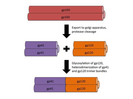
<br>The envelope of the HIV virion consists of a glycoprotein complex, called Env, embedded in a host-sourced phospholipid membrane. Each virion includes approximately 15 Env glycoprotein complexes [6]. Env itself consists of trimers of noncovalently bound gp120 and gp41 subunits. During replication, the integrated prophage is transcribed, producing Env mRNA that is read by endoplasmic reticulum ribosomes to produce an 845-870 amino acid precursor polypeptide. This precursor is modified with the addition of asparagine linked, high mannose content sugar chains, yielding the intermediate glycoprotein gp160 [12]. The gp160 glycoprotein forms homotrimers before being exported to the golgi apparatus, where host proteases digest the glycoprotein complex, yielding gp120 and gp41 subunits which remain as homotrimers. The gp120 and gp41 trimer bundles are further modified through N-glycosylation. This glycosylation step alone contributes a great deal to the variability of the Env protein structure; gp120, for example, has around 24 potential N-glycosylation sites allowing for a wide variety of possible N-glycosylation combinations [7, 12]. | <br>The envelope of the HIV virion consists of a glycoprotein complex, called Env, embedded in a host-sourced phospholipid membrane. Each virion includes approximately 15 Env glycoprotein complexes [6]. Env itself consists of trimers of noncovalently bound gp120 and gp41 subunits. During replication, the integrated prophage is transcribed, producing Env mRNA that is read by endoplasmic reticulum ribosomes to produce an 845-870 amino acid precursor polypeptide. This precursor is modified with the addition of asparagine linked, high mannose content sugar chains, yielding the intermediate glycoprotein gp160 [12]. The gp160 glycoprotein forms homotrimers before being exported to the golgi apparatus, where host proteases digest the glycoprotein complex, yielding gp120 and gp41 subunits which remain as homotrimers. The gp120 and gp41 trimer bundles are further modified through N-glycosylation. This glycosylation step alone contributes a great deal to the variability of the Env protein structure; gp120, for example, has around 24 potential N-glycosylation sites allowing for a wide variety of possible N-glycosylation combinations [7, 12]. | ||
After N-glycosylation, the gp120 trimer bundles form noncovalent bonds with gp41 trimer bundles, yielding gp41-gp120 bundle heterodimers (figure 2). Finally, the mature Env proteins are sent to the cell membrane and are embedded in virions budding from the cell. The high mutation rate of HIV means that mutations to these Env components are fairly common, resulting in a large number of progeny virions with Env glycoprotein complexes that fail to mature properly or simply fall out of the viral envelope [12]. Virions with these non-viable Env protein complexes are rendered incapable of infection. The sheer number of virions produced, however, guarantees that at least a small portion of the progeny virions will have viable Env protein complexes. | After N-glycosylation, the gp120 trimer bundles form noncovalent bonds with gp41 trimer bundles, yielding gp41-gp120 bundle heterodimers (figure 2). Finally, the mature Env proteins are sent to the cell membrane and are embedded in virions budding from the cell. The high mutation rate of HIV means that mutations to these Env components are fairly common, resulting in a large number of progeny virions with Env glycoprotein complexes that fail to mature properly or simply fall out of the viral envelope [12]. Virions with these non-viable Env protein complexes are rendered incapable of infection. The sheer number of virions produced, however, guarantees that at least a small portion of the progeny virions will have viable Env protein complexes. | ||
<br> | <br> | ||
==Section 2== | ==Section 2== | ||
Revision as of 02:34, 23 April 2014
Introduction
By Ian Perrone
HIV, or human immunodeficiency virus, is an enveloped sense-RNA retrovirus that infects human immune cells in order to replicate (figure 1).
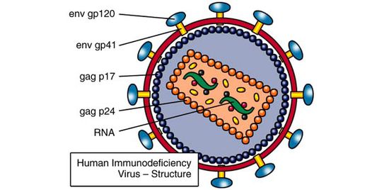
Its classification as a retrovirus is due to its use of reverse transcriptase to copy its RNA genome into DNA. This DNA is imported into the host cell nucleus, where it is inserted into the host cell genome by virally encoded integrase. At this point, the virus lives as a prophage, sporadically producing infective virions through use of host transcriptase and ribosomes. Specifically, HIV is known as a lentivirus, a form of retrovirus with a long incubation time due to relatively slow replication. Despite this relatively slow replication speed, unless treated with HAART (highly active anti-retroviral therapy), the vast majority of HIV patients will eventually develop AIDS (acquired immunodeficiency syndrome). AIDS is characterized by extremely heightened susceptibility to opportunistic infection by rarely pathogenic bacteria, such as Pneumocystis carinii and the development of uncommon types of cancer, such as Kaposi sarcoma [6]. Since the original spread of the virus from Africa to the USA in 1981, HIV and AIDS have caused the deaths of over 25 million people across the globe [6].
Like all enveloped viruses, HIV has envelope proteins that recognize particular protein structures on target cells. These envelope proteins are what allow viruses to bind to cells before fusing with the cell membrane entering the cytoplasm. These proteins also define the viral tropism, or range of cells that the virus can infect, making knowledge of the viral envelope proteins necessary for understanding the pathogenicity of the virus [2]. In addition, the key role played by the envelope proteins in infecting target cells means that a wide variety of treatments for viral diseases have been designed to block the action of these envelope proteins. The design of these binding/fusion blockers also requires a firm grasp of viral envelope proteins, especially in HIV, which has an especially high mutation rate due to its use of relatively inaccurate reverse transcriptase for replication. Below, we explore the structural characteristics of the main HIV envelope protein components, gp120 and gp41, as well as their respective targets. We also examine the influence these proteins have on viral evolution patterns, with a specific focus on the evolution of viral quasispecies within individual patients. Finally, we consider the potential for HIV treatments targeting these envelope proteins, as well as the difficulties associated with treatment of such a mutation-prone virus.
At right is a sample image insertion. It works for any image uploaded anywhere to MicrobeWiki. The insertion code consists of:
Double brackets: [[
Filename: PHIL_1181_lores.jpg
Thumbnail status: |thumb|
Pixel size: |300px|
Placement on page: |right|
Legend/credit: Electron micrograph of the Ebola Zaire virus. This was the first photo ever taken of the virus, on 10/13/1976. By Dr. F.A. Murphy, now at U.C. Davis, then at the CDC.
Closed double brackets: ]]
Other examples:
Bold
Italic
Subscript: H2O
Superscript: Fe3+
Introduce the topic of your paper. What microorganisms are of interest? Habitat? Applications for medicine and/or environment?
[[File:|thumb||550px||right|Figure X. ]]
[[File:|thumb||550px||right|Figure X. ]]
[[File:|thumb||550px||right|Figure X. ]]
HIV Env Overview
The envelope of the HIV virion consists of a glycoprotein complex, called Env, embedded in a host-sourced phospholipid membrane. Each virion includes approximately 15 Env glycoprotein complexes [6]. Env itself consists of trimers of noncovalently bound gp120 and gp41 subunits. During replication, the integrated prophage is transcribed, producing Env mRNA that is read by endoplasmic reticulum ribosomes to produce an 845-870 amino acid precursor polypeptide. This precursor is modified with the addition of asparagine linked, high mannose content sugar chains, yielding the intermediate glycoprotein gp160 [12]. The gp160 glycoprotein forms homotrimers before being exported to the golgi apparatus, where host proteases digest the glycoprotein complex, yielding gp120 and gp41 subunits which remain as homotrimers. The gp120 and gp41 trimer bundles are further modified through N-glycosylation. This glycosylation step alone contributes a great deal to the variability of the Env protein structure; gp120, for example, has around 24 potential N-glycosylation sites allowing for a wide variety of possible N-glycosylation combinations [7, 12].
After N-glycosylation, the gp120 trimer bundles form noncovalent bonds with gp41 trimer bundles, yielding gp41-gp120 bundle heterodimers (figure 2). Finally, the mature Env proteins are sent to the cell membrane and are embedded in virions budding from the cell. The high mutation rate of HIV means that mutations to these Env components are fairly common, resulting in a large number of progeny virions with Env glycoprotein complexes that fail to mature properly or simply fall out of the viral envelope [12]. Virions with these non-viable Env protein complexes are rendered incapable of infection. The sheer number of virions produced, however, guarantees that at least a small portion of the progeny virions will have viable Env protein complexes.

