Peroxisome Protein Import in S.cerevisiae: Difference between revisions
(Created page with "by Charlotte Leblang") |
No edit summary |
||
| Line 1: | Line 1: | ||
==Introduction== | |||
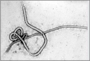
[[Image:Ebola_virus2.jpg|thumb|300px|right|Electron micrograph of the Ebola Zaire virus. This was one of the first micrographs taken of the virus, in 1976. By Dr. Frederick Murphy, now at U.C. Davis, then at the [http://wwwnc.cdc.gov/eid/article/21/11/pdfs/et-2111.pdf CDC].]] | |||
<br>By Charlotte Leblang<br> | |||
<br>At right is a sample image insertion. It works for any image uploaded anywhere to MicrobeWiki. The insertion code consists of: | |||
<br><b>Double brackets:</b> [[ | |||
<br><b>Filename:</b> Ebola_virus2.jpg | |||
<br><b>Thumbnail status:</b> |thumb| | |||
<br><b>Pixel size:</b> |300px| | |||
<br><b>Placement on page:</b> |right| | |||
<br><b>Legend/credit:</b> Electron micrograph of the Ebola Zaire virus. This was the first photo ever taken of the virus, on 10/13/1976. By Dr. F.A. Murphy, now at U.C. Davis, then at the [http://wwwnc.cdc.gov/eid/article/21/11/pdfs/et-2111.pdf CDC]. | |||
<br><b>Closed double brackets:</b> ]] | |||
<br><br>Other examples: | |||
<br><b>Bold</b> | |||
<br><i>Italic</i> | |||
<br><b>Subscript:</b> H<sub>2</sub>O | |||
<br><b>Superscript:</b> Fe<sup>3+</sup> | |||
<br>Introduce the topic of your paper. State your health service question, and explain the biomedical issues.<br> | |||
==Section 1== | |||
Include some current research, with at least one figure showing data.<br> | |||
<br> | |||
==Section 2== | |||
Include some current research, with at least one figure showing data.<br> | |||
<br> | |||
==Section 3== | |||
Include some current research, with at least one figure showing data.<br> | |||
<br> | |||
==Conclusion== | |||
<br><br> | |||
==References== | |||
[1] [http://www.plosbiology.org/article/fetchObject.action?uri=info%3Adoi%2F10.1371%2Fjournal.pbio.1000005&representation=PDF Hodgkin, J. and Partridge, F.A. "<i>Caenorhabditis elegans</i> meets microsporidia: the nematode killers from Paris." 2008. PLoS Biology 6:2634-2637.] | |||
<br><br>Authored for BIOL 291.00 Health Service and Biomedical Analysis, taught by [mailto:slonczewski@kenyon.edu Joan Slonczewski], 2016, [http://www.kenyon.edu/index.xml Kenyon College]. | |||
Revision as of 18:57, 26 October 2015
Introduction

By Charlotte Leblang
At right is a sample image insertion. It works for any image uploaded anywhere to MicrobeWiki. The insertion code consists of:
Double brackets: [[
Filename: Ebola_virus2.jpg
Thumbnail status: |thumb|
Pixel size: |300px|
Placement on page: |right|
Legend/credit: Electron micrograph of the Ebola Zaire virus. This was the first photo ever taken of the virus, on 10/13/1976. By Dr. F.A. Murphy, now at U.C. Davis, then at the CDC.
Closed double brackets: ]]
Other examples:
Bold
Italic
Subscript: H2O
Superscript: Fe3+
Introduce the topic of your paper. State your health service question, and explain the biomedical issues.
Section 1
Include some current research, with at least one figure showing data.
Section 2
Include some current research, with at least one figure showing data.
Section 3
Include some current research, with at least one figure showing data.
Conclusion
References
Authored for BIOL 291.00 Health Service and Biomedical Analysis, taught by Joan Slonczewski, 2016, Kenyon College.
