Quorum Sensing in Methanosaeta harundinacea 6Ac: Difference between revisions
No edit summary |
No edit summary |
||
| Line 10: | Line 10: | ||
[http://stke.sciencemag.org.ezproxy.library.ubc.ca/cgi/reprint/pnas;99/5/3129.pdf Quorum sensing related virulence in ''Vibrio cholerae''] | [http://stke.sciencemag.org.ezproxy.library.ubc.ca/cgi/reprint/pnas;99/5/3129.pdf Quorum sensing related virulence in ''Vibrio cholerae''] | ||
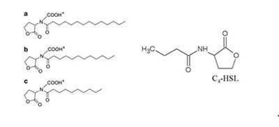
[[File:Autoinducers.jpeg|400px|thumb|right| Left: the 3 different carboxyl-AHLs used by M. harundinacea 6Ac. Right: acyl homoserine lactone (AHL), the autoinducer used by Gram negative bacteria. C4-HSL is shown.]] | |||
==Autoinducers in ''M. harundinacea'' 6Ac== | ==Autoinducers in ''M. harundinacea'' 6Ac== | ||
The autoinducers used by ''M. harundinacea'' 6Ac are carboxyl-AHLs, which are essentially AHLs with a carboxyl moiety attached to the nitrogen (Zhang et al. 2012). There are 3 slight variants synthesized (see figure 1), which are N-carboxyl-C14-HSL (homoserine lactone), N-carboxyl-C12-HSL, and N-carboxyl-C10-HSL (Zhang et al. 2012). These are made by the enzyme AHL synthase, encoded for by the ''fliI'' gene, which is basally transcribed(Zhang et al. 2012). The sensor protein for the carboxyl-AHLs, FilR, is coded for by the ''filR'' gene which is also basally transcribed, and the protein binds the autoinducers when they reach a threshold concentration (Zhang et al. 2012), the concentration which is an indication of increased cell-density in the area. The FilR-autoinducer complex binds to certain genes in the DNA resulting in up-regulation of their transcription, and down-regulation of other genes, which results in changes in physiology of the cells as well as a different metabolic pattern (Zhang et al. 2012). | The autoinducers used by ''M. harundinacea'' 6Ac are carboxyl-AHLs, which are essentially AHLs with a carboxyl moiety attached to the nitrogen (Zhang et al. 2012). There are 3 slight variants synthesized (see figure 1), which are N-carboxyl-C14-HSL (homoserine lactone), N-carboxyl-C12-HSL, and N-carboxyl-C10-HSL (Zhang et al. 2012). These are made by the enzyme AHL synthase, encoded for by the ''fliI'' gene, which is basally transcribed(Zhang et al. 2012). The sensor protein for the carboxyl-AHLs, FilR, is coded for by the ''filR'' gene which is also basally transcribed, and the protein binds the autoinducers when they reach a threshold concentration (Zhang et al. 2012), the concentration which is an indication of increased cell-density in the area. The FilR-autoinducer complex binds to certain genes in the DNA resulting in up-regulation of their transcription, and down-regulation of other genes, which results in changes in physiology of the cells as well as a different metabolic pattern (Zhang et al. 2012). | ||
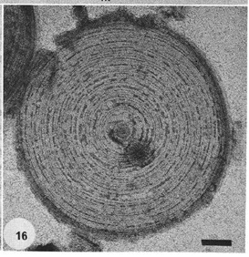
[[File: | [[File:EM spacer plug.jpeg|300px|thumb|left| Negatively stained EM micrograph depicting a spacer plug which has been separated from the filament. The plug is firmly attached to the sheath, and is composed of numerous concentric rings.]] | ||
==Changes in Physiology== | ==Changes in Physiology== | ||
When the cell density is low, ''M. harundinacea'' 6Ac exist as short, singular rod-shaped cells (Ma et al. 2006), but when the cell density is high the archaeon’s physiology drastically changes. Chains of cells are stacked end to end forming filaments that can be greater than 200 microns, in contrast to a length of 3-5 microns as single cells (Guo et al. 2009). Beveridge et al. (1986) found that the filaments consist of four major parts: the cells lined up in a chain, spacer plugs which separated each individual cell as well as plugging the two ends of the filament, an amorphous granular matrix, and encasing everything a proteinaceous sheath. The spacer plugs are anchored to the sheath, and appear to have multiple layers. The granular matrix is enclosed by the sheath and the plugs and completely encloses individual cells. The protein sheath is striated and composed of a highly crystalline structure. This lends it tremendous strength against pressure, as well as other physical and chemical disruption (Beveridge et al. 1986). As such, when ''M. harundinacea'' 6Ac form into filaments, they can better withstand unfavourable environment conditions and thus have an edge over other micro-organisms in the area. Note that Beveridge et al. (1986) described filaments formed by ''Methanothrix concilii'', which have been renamed to ''Methanosaeta concilii'' as elucidated by Patel and Sprott (1990). Given that there is a 92.5% similarity between ''M. concilii'' and ''M. harundinacea'' 6Ac based on 16S rRNA sequences (Ma et al. 2006), the filament descriptions are likely accurate for ''M. harundinacea'' 6Ac as well. | When the cell density is low, ''M. harundinacea'' 6Ac exist as short, singular rod-shaped cells (Ma et al. 2006), but when the cell density is high the archaeon’s physiology drastically changes. Chains of cells are stacked end to end forming filaments that can be greater than 200 microns, in contrast to a length of 3-5 microns as single cells (Guo et al. 2009). Beveridge et al. (1986) found that the filaments consist of four major parts: the cells lined up in a chain, spacer plugs which separated each individual cell as well as plugging the two ends of the filament, an amorphous granular matrix, and encasing everything a proteinaceous sheath. The spacer plugs are anchored to the sheath, and appear to have multiple layers. The granular matrix is enclosed by the sheath and the plugs and completely encloses individual cells. The protein sheath is striated and composed of a highly crystalline structure. This lends it tremendous strength against pressure, as well as other physical and chemical disruption (Beveridge et al. 1986). As such, when ''M. harundinacea'' 6Ac form into filaments, they can better withstand unfavourable environment conditions and thus have an edge over other micro-organisms in the area. Note that Beveridge et al. (1986) described filaments formed by ''Methanothrix concilii'', which have been renamed to ''Methanosaeta concilii'' as elucidated by Patel and Sprott (1990). Given that there is a 92.5% similarity between ''M. concilii'' and ''M. harundinacea'' 6Ac based on 16S rRNA sequences (Ma et al. 2006), the filament descriptions are likely accurate for ''M. harundinacea'' 6Ac as well. | ||
[[File:Filament segment.jpeg|600px|thumb| | [[File:Filament segment.jpeg|600px|thumb|right| A segment of the filament (3 individual cells) formed by M. harundinacea. The four major components of the filaments, the outer protein sheath, the spacer plugs, granular matrix, and the individual archaea are shown.]] | ||
== | ==Changes in Metabolism== | ||
The | The metabolic patterns between the 2 distinct morphologies are significantly different. Both methane production and the reaction rate of methanogenesis are higher in the filamentous cells than in the short rods (Zhang et al. 2012). It was also shown by Zhang et al. (2012) that the filamentous cells produce lower levels of cellular proteins compared to the short cells, and had less biomass. The significance of this is that ''M. harundinacea'' 6Ac can better compete with other micro-organisms for resources in the environment, such as for acetate with acetoclastic microbes, and as well they can divide quicker due to faster metabolism and the lower biomass required, and outgrow any competitors. | ||
=Metabolism= | =Metabolism= | ||
Revision as of 22:13, 14 December 2012
Overview
The archaeon Methanosaeta harundinacea 6Ac is part of a family of aceticlastic methanogens who exclusively catabolize acetate (Ferry 2008). It is rod-shaped, non-motile, and usually found singly or in pairs, but when grown in acetate with high cell density, the cells form into filaments which are self-contained (Ma et. al 2006), products of quorum sensing.
Quorum Sensing
Quorum sensing is a cell density-dependent process of gene regulation by which unicellular organisms can communicate with others of the same species and co-ordinate their behaviours (Miller and Bassler 2001). Signal molecules called autoinducers are constitutively expressed and secreted out of the cell, which then get taken up by members of the other species present in the area. As such, autoinducer concentration in the immediate environment increases as cell density increases and given that enough autoinducers are uptaken, a threshold concentration is reached inside the cells which triggers a change in gene expression. The cells can then co-ordinate a number of processes together, such as biofilm formation, sporulation (Miller and Bassler 2001, production of virulence factors such as in Pseudomonas aeruginosa (Pesci et al. 1997) , or in the case of Vibrio fischeri, bioluminescence (Hastings and Greenberg 1999). In Gram-positive bacteria, oligopeptides serve as autoinducers, and Gram-negative bacteria use acyl homoserine lactones (AHLs) (Miller and Bassler 2001).
More on quorum sensing
Quorum sensing related virulence in Vibrio cholerae
Autoinducers in M. harundinacea 6Ac
The autoinducers used by M. harundinacea 6Ac are carboxyl-AHLs, which are essentially AHLs with a carboxyl moiety attached to the nitrogen (Zhang et al. 2012). There are 3 slight variants synthesized (see figure 1), which are N-carboxyl-C14-HSL (homoserine lactone), N-carboxyl-C12-HSL, and N-carboxyl-C10-HSL (Zhang et al. 2012). These are made by the enzyme AHL synthase, encoded for by the fliI gene, which is basally transcribed(Zhang et al. 2012). The sensor protein for the carboxyl-AHLs, FilR, is coded for by the filR gene which is also basally transcribed, and the protein binds the autoinducers when they reach a threshold concentration (Zhang et al. 2012), the concentration which is an indication of increased cell-density in the area. The FilR-autoinducer complex binds to certain genes in the DNA resulting in up-regulation of their transcription, and down-regulation of other genes, which results in changes in physiology of the cells as well as a different metabolic pattern (Zhang et al. 2012).
Changes in Physiology
When the cell density is low, M. harundinacea 6Ac exist as short, singular rod-shaped cells (Ma et al. 2006), but when the cell density is high the archaeon’s physiology drastically changes. Chains of cells are stacked end to end forming filaments that can be greater than 200 microns, in contrast to a length of 3-5 microns as single cells (Guo et al. 2009). Beveridge et al. (1986) found that the filaments consist of four major parts: the cells lined up in a chain, spacer plugs which separated each individual cell as well as plugging the two ends of the filament, an amorphous granular matrix, and encasing everything a proteinaceous sheath. The spacer plugs are anchored to the sheath, and appear to have multiple layers. The granular matrix is enclosed by the sheath and the plugs and completely encloses individual cells. The protein sheath is striated and composed of a highly crystalline structure. This lends it tremendous strength against pressure, as well as other physical and chemical disruption (Beveridge et al. 1986). As such, when M. harundinacea 6Ac form into filaments, they can better withstand unfavourable environment conditions and thus have an edge over other micro-organisms in the area. Note that Beveridge et al. (1986) described filaments formed by Methanothrix concilii, which have been renamed to Methanosaeta concilii as elucidated by Patel and Sprott (1990). Given that there is a 92.5% similarity between M. concilii and M. harundinacea 6Ac based on 16S rRNA sequences (Ma et al. 2006), the filament descriptions are likely accurate for M. harundinacea 6Ac as well.
Changes in Metabolism
The metabolic patterns between the 2 distinct morphologies are significantly different. Both methane production and the reaction rate of methanogenesis are higher in the filamentous cells than in the short rods (Zhang et al. 2012). It was also shown by Zhang et al. (2012) that the filamentous cells produce lower levels of cellular proteins compared to the short cells, and had less biomass. The significance of this is that M. harundinacea 6Ac can better compete with other micro-organisms for resources in the environment, such as for acetate with acetoclastic microbes, and as well they can divide quicker due to faster metabolism and the lower biomass required, and outgrow any competitors.
Metabolism
Ignicoccus species are chemolithoautotrophs that use molecular hydrogen as the inorganic electron donor and elemental sulphur as the inorganic terminal electron acceptor[1] . The reduction of the elemental sulphur results in the production of hydrogen sulphide gas.
Ignicoccus are autotrophs in that they fix their own carbon dioxide into organic molecules. The carbon dioxide fixation process they use is a novel process called a dicarboxylate/4-hydroxybutyrate autotrophic carbon assimilation cycle that involves 14 different enzymes[8] .
Members of the Ignicoccus genus are able to use ammonium as a nitrogen source.
Growth Conditions
Because members of the Ignicoccus genus are hyperthermophiles and obligate anaerobes, it is not surprising that their growth conditions are very complex. They are grown in a liquid medium known as ½ SME Ignicoccus which is a solution of synthetic sea water which is then made anaerobic.
Grown in this media at their optimal growth temperature of 90C, the members of the Ignicoccus genus typically reach a cell density of ~4x10⁷cells/mL[1] .
The addition of yeast extract to the ½ SME media has been shown to stimulate the growth and increase maximum cell density achieved. The mechanism by which this is achieved is not known[1] .
Symbiosis
Ignicoccus hospitalis is the only member of the genus Ignicoccus that has been shown to have an extensive symbiotic relationship with another organism.
Ignicoccus hospitalis has been shown to engage in symbiosis with Nanoarchaeum equitans . Nanoarchaeum equitans is a very small coccoid species with a cell diameter of 0.4 µm[9] . Genome analysis has provided much of the known information about this species.
To further complicate the symbiotic relationship between both species, it’s been observed that the presence of Nanoarchaeum equitans on the surface of Ignicoccus hospitalis somehow inhibits the cell replication of Ignicoccus hospitalis . How or why this occurs has not yet been elucidated[3] .

Nanoarchaeum equitans
Nanoarchaeum equitans has the smallest non-viral genome ever sequenced at 491kb[9] . Analysis of the genome sequence indicates that 95% of the predicted proteins and stable RNA molecules are somehow involved in repair and replication of the cell and its genome[3] .
Analysis of the genome also showed that Nanoarchaeum equitans lacks nearly all genes known to be required in amino acid, nucleotide, cofactor and lipid metabolism. This is partially supported by the evidence that Nanoarchaeum equitans has been shown to derive its cell membrane from its host Ignicoccus hospitalis cell membrane. The direct contact observed between Nanoarchaeum equitans and Ignicoccus hospitalis is hypothesized to form a pore between the two organisms in order to exchange metabolites or substrates (likely from Ignicoccus hospitalis towards Nanoarchaeum equitans due to the parasitic relationship). The exchange of periplasmic vesicles is not thought to be involved in metabolite or substrate exchange despite the presence of vesicles in the periplasm of Ignicoccus hospitalis .
These analyses of the Nanoarchaeum equitans genome support the fact of the extensive symbiotic relationship between Nanoarchaeum equitans and Ignicoccus hospitalis. However, it has not yet been proven that it is a strictly parasitic relationship and further research may prove that there is a commensal relationship between the two species.
References
(1) Burggraf S., Huber H., Mayer T., Rachel R., Stetter K.O. and Wyschkony I. ” Ignicoccus gen. nov., a novel genus of hyperthermophilic, chemolithoautotrophic Archaea, represented by two new species, Ignicoccus islandicus sp. nov. and Ignicoccus pacificus sp. nov.” International Journal of Systematic and Evolutionary Microbiology, 2000, Volume 50.
(2) Naether D.J. and Rachel R. “The outer membrane of the hyperthermophilic archaeon Ignicoccus: dynamics, ultrastructure and composition.” Biochemical Society Transactions, 2004, Volume 32, part 2.
(3) Giannone R.J., Heimerl T., Hettich R.L., Huber H., Karpinets T., Keller M., Kueper U., Podar M. and Rachel R. “Proteomic Characterization of Cellular and Molecular Processes that Enable the Nanoarchaeum equitans- Ignicoccus hospitalis Relationship.” PLoS ONE, 2011, Volume 6, Issue 8.
(4) Eisenreich W., Gallenberger M., Huber H., Jahn U., Junglas B., Paper W., Rachel R. and Stetter K.O. “Nanoarchaeum equitans and Ignicoccus hospitalis: New Insights into a Unique, Intimate Association of Two Archaea.” Journal of Bacteriology, 2008, DOI: 10.1128/JB.01731-07.
(5) Grosjean E., Huber H., Jahn U., Sturt H, and Summons R. “Composition of the lipids of Nanoarchaeum equitans and their origin from its host Ignicoccus sp. strain KIN4/I.” Arch Microbiol, 2004, DOI: 10.1007/s00203-004-0725-x.
(6) Briegel A., Burghardt T., Huber H., Junglas B., Rachel R., Walther P. and Wirth R. “Ignicoccus hospitalis and Nanoarchaeum equitans: ultrastructure, cell–cell interaction, and 3D reconstruction from serial sections of freeze-substituted cells and by electron cryotomography.” Arch Microbiol, 2008, DOI 10.1007/s00203-008-0402-6.
(7) Burghardt T., Huber H., Junglas B., Naether D.J. and Rachel R. “The dominating outer membrane protein of the hyperthermophilic Archaeum Ignicoccus hospitalis: a novel pore-forming complex.” Molecular Microbiology, 2007, Volume 63.
(8) Berg I.A., Eisenreich W., Eylert E., Fuchs G., Gallenberger M., Huber H.,Jahn U. and Kockelkorn D. “A dicarboxylate/4-hydroxybutyrate autotrophic carbon assimilation cycle in the hyperthermophilic Archaeum Ignicoccus hospitalis.” PNAS, 2008, Volume 105, issue 22.
(9) Brochier C., Gribaldo S., Zivanovic Y., Confalonieri F. and Forterre P. “Nanoarchaea: representatives of a novel archaeal phylum or a fast-evolving euryarchaeal lineage related to Thermococcales?” Genome Biology 2005, DOI:10.1186/gb-2005-6-5-r42.
(10) Huber H., Rachel R., Riehl S. and Wyschkony I. “The ultrastructure of Ignicoccus: Evidence for a novel outer membrane and for intracellular vesicle budding in an archaeon.” Archaea, 2002, Volume 1.


