Bacillus Anthracis: Anthrax Lethal Toxin: Difference between revisions
(Created page with "==Introduction== <br>By Connor Gibbons<br> <br>At right is a sample image insertion. It works for any image uploaded anywhere to MicrobeWiki. The insertion code consists of:...") |
No edit summary |
||
| Line 16: | Line 16: | ||
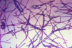
[[Image:Bacillus_anthracis_gram.jpg|thumb|300px|center|A photomicrograph of Bacillus anthracis bacteria using Gram-stain technique. Photo obtained from the CDC's Public Health Image Library.]] | [[Image:Bacillus_anthracis_gram.jpg|thumb|300px|center|A photomicrograph of Bacillus anthracis bacteria using Gram-stain technique. Photo obtained from the CDC's Public Health Image Library.]] | ||
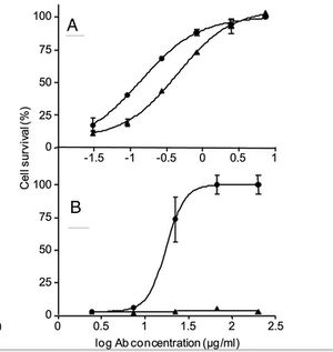
[[Image:cAb29Fig.jpg|thumb|300px|center|Figure 1. A and B show the neutralization assay performed using cultured J774A macrophages that were incubated for 5 h with fixed amounts of LF and PA83 (A) or purified prepore (B) in the presence of cAb29 (circles) or Ab33 (triangles) in the indicated concentrations. Cell survival was determined by XTT and plotted as the percentage of untreated control cells. Points are mean ± S.D. of triplicate determinants. Ab concentration, antibody concentration.]] | |||
<br>Introduce the topic of your paper. What microorganisms are of interest? Habitat? Applications for medicine and/or environment?<br> | <br>Introduce the topic of your paper. What microorganisms are of interest? Habitat? Applications for medicine and/or environment?<br> | ||
Revision as of 11:43, 23 April 2013
Introduction
By Connor Gibbons
At right is a sample image insertion. It works for any image uploaded anywhere to MicrobeWiki. The insertion code consists of:
Double brackets: [[
Filename: PHIL_1181_lores.jpg
Thumbnail status: |thumb|
Pixel size: |300px|
Placement on page: |right|
Legend/credit: Electron micrograph of the Ebola Zaire virus. This was the first photo ever taken of the virus, on 10/13/1976. By Dr. F.A. Murphy, now at U.C. Davis, then at the CDC.
Closed double brackets: ]]
Other examples:
Bold
Italic
Subscript: H2O
Superscript: Fe3+

Introduce the topic of your paper. What microorganisms are of interest? Habitat? Applications for medicine and/or environment?
Section 1
Include some current research, with at least one figure showing data.
Section 2
Include some current research, with at least one figure showing data.
Section 3
Include some current research, with at least one figure showing data.
Conclusion
Overall text length at least 3,000 words, with at least 3 figures.
References
Edited by student of Joan Slonczewski for BIOL 238 Microbiology, 2011, Kenyon College.

