Bacillus Anthracis: Anthrax Lethal Toxin: Difference between revisions
No edit summary |
No edit summary |
||
| Line 17: | Line 17: | ||
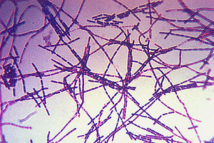
[[Image:Bacillus_anthracis_gram.jpg|thumb|300px|center|A photomicrograph of Bacillus anthracis bacteria using Gram-stain technique. Photo obtained from the CDC's Public Health Image Library.]] | [[Image:Bacillus_anthracis_gram.jpg|thumb|300px|center|A photomicrograph of Bacillus anthracis bacteria using Gram-stain technique. Photo obtained from the CDC's Public Health Image Library.]] | ||
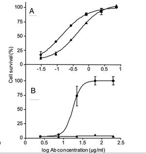
[[Image:cAb29Fig.jpg|thumb|300px|center|Figure 1. A and B show the neutralization assay performed using cultured J774A macrophages that were incubated for 5 h with fixed amounts of LF and PA83 (A) or purified prepore (B) in the presence of cAb29 (circles) or Ab33 (triangles) in the indicated concentrations. Cell survival was determined by XTT and plotted as the percentage of untreated control cells. Points are mean ± S.D. of triplicate determinants. Ab concentration, antibody concentration.]] | [[Image:cAb29Fig.jpg|thumb|300px|center|Figure 1. A and B show the neutralization assay performed using cultured J774A macrophages that were incubated for 5 h with fixed amounts of LF and PA83 (A) or purified prepore (B) in the presence of cAb29 (circles) or Ab33 (triangles) in the indicated concentrations. Cell survival was determined by XTT and plotted as the percentage of untreated control cells. Points are mean ± S.D. of triplicate determinants. Ab concentration, antibody concentration (Mechaly).]] | ||
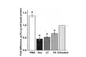
[[Image:surfactantFig.png|thumb|300px|center|LT decreases surfactant production in AEC. Early-culture AEC were treated with LT (2�g/ml) for 72 h and harvested. Phorbol 12-myristate (PMA; 50 ng/ml) and dexamethasone (Dex; 1�M) were used as positive and negative controls, respectively. Surfactant levels in supernatants were measured by a phospholipid assay and normalized to protein content per sample. Data are means SEM of the results from 3 experiments on 3 separate individuals, with 2 to 4 trials for each treatment. *, P value of �0.05 versus untreated cells.]] | [[Image:surfactantFig.png|thumb|300px|center|LT decreases surfactant production in AEC. Early-culture AEC were treated with LT (2�g/ml) for 72 h and harvested. Phorbol 12-myristate (PMA; 50 ng/ml) and dexamethasone (Dex; 1�M) were used as positive and negative controls, respectively. Surfactant levels in supernatants were measured by a phospholipid assay and normalized to protein content per sample. Data are means SEM of the results from 3 experiments on 3 separate individuals, with 2 to 4 trials for each treatment. *, P value of �0.05 versus untreated cells (Langer).]] | ||
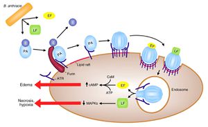
[[Image:BAnthracisPathogenesis.jpg|thumb|300px|center|The process of <i>B. anthracis</i> pathogenesis begins with the binding of PA to a host cell. The PA molecule is then cleaved and oligomerizes, at which time it can bind LF and EF forming LeTx and EdTx. These complexes are then internalized into the cell, where LeTx causes reduced intracellular MAPK concentration, resulting in necrosis and hypoxia, and EdTx causes increased intracellular cAMP concentration, resulting in edema around the infection site.]] | |||
Revision as of 23:39, 24 April 2013
Introduction
By Connor Gibbons
At right is a sample image insertion. It works for any image uploaded anywhere to MicrobeWiki. The insertion code consists of:
Double brackets: [[
Filename: PHIL_1181_lores.jpg
Thumbnail status: |thumb|
Pixel size: |300px|
Placement on page: |right|
Legend/credit: Electron micrograph of the Ebola Zaire virus. This was the first photo ever taken of the virus, on 10/13/1976. By Dr. F.A. Murphy, now at U.C. Davis, then at the CDC.
Closed double brackets: ]]
Other examples:
Bold
Italic
Subscript: H2O
Superscript: Fe3+


Introduce the topic of your paper. What microorganisms are of interest? Habitat? Applications for medicine and/or environment?
Section 1
Include some current research, with at least one figure showing data.
Section 2
Include some current research, with at least one figure showing data.
Section 3
Include some current research, with at least one figure showing data.
Conclusion
Overall text length at least 3,000 words, with at least 3 figures.
References
Edited by student of Joan Slonczewski for BIOL 238 Microbiology, 2011, Kenyon College.

