Marburgvirus: Difference between revisions
(Corrected claimed magnification of image to match that of source. But see my comments that I will add in the discussion page…) |
|||
| (12 intermediate revisions by 2 users not shown) | |||
| Line 1: | Line 1: | ||
{{Uncurated}} | |||
==Classification== | ==Classification== | ||
Viruses | Viruses; ssRNA negative-strand viruses; Mononegavirales; Filoviridae | ||
===Species=== | ===Species=== | ||
| Line 8: | Line 9: | ||
==Description and Significance== | ==Description and Significance== | ||
Marburg is an extremely dangerous virus as it has a human fatality rate of 25-90%, with outbreaks that are impossible to predict or prevent.[1][7] Marburgvirus was the first ever filovirus recognized, discovered in 1967 when laboratory workers in Marburg, Germany developed hemorrhagic fever after handling tissue of African green monkeys from Uganda. The 1967 outbreak proved fatal in seven of the 37 cases. | Marburg is an extremely dangerous virus as it has a human fatality rate of 25-90%, with outbreaks that are impossible to predict or prevent.[1][7] Marburgvirus was the first ever filovirus recognized, discovered in 1967 when laboratory workers in Marburg, Germany developed hemorrhagic fever after handling tissue of African green monkeys from Uganda. The 1967 outbreak proved fatal in seven of the 37 cases. | ||
In 1975 the virus reemerged in Johannesburg, South Africa in a person who had been traveling in Zimbabwe. Several other cases of Marburg occurred sporadically through 1980, all of patients who had been recently traveling in Africa. In 1998, an outbreak occurred in Congo amongst individuals who had been working in a gold mine. In all cases, attempts to isolate the virus from all locations were unsuccessful. The virus has been appearing sporadically throughout parts of Africa since. In Angola from October 2004 to August 2005 for instance, an outbreak of Marburg occurred leaving 329 of 374 infected people dead (88% mortality). Cases of Marburgvirus continue to be rare and random, though most recently occurring in 2008 in Colorado of a man who had been traveling in Uganda.[7] | |||
The only other filovirus is the Ebola virus, which claims an even higher mortality rate, discovered after Marburg in 1976.[3] The natural origins of both | In 1975 the virus reemerged in Johannesburg, South Africa in a person who had been traveling in Zimbabwe. Several other cases of Marburg occurred sporadically through 1980, all of patients who had been recently traveling in Africa. In 1998, an outbreak occurred in Congo amongst individuals who had been working in a gold mine. In all cases, attempts to isolate the virus from all locations were unsuccessful. The virus has been appearing sporadically throughout parts of Africa since. In Angola from October 2004 to August 2005 for instance, an outbreak of Marburg occurred leaving 329 of 374 infected people dead (88% mortality). | ||
Cases of Marburgvirus continue to be rare and random, though most recently occurring in 2008 in Colorado of a man who had been traveling in Uganda.[7] | |||
The only other filovirus is the Ebola virus, which claims an even higher mortality rate, discovered after Marburg in 1976.[3] The natural origins of both remain a complete mystery to this day and a cause of major concern and research.[1] | |||
==Genome Structure== | ==Genome Structure== | ||
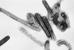
[[Image:Marburg2.jpg|frame|Negative stain image of an isolate of Marburg virus, | [[Image:Marburg2.jpg|frame|Negative stain image of an isolate of Marburg virus, 100,000x magnification. Note filamentous particles [4]. ]] | ||
Filoviruses are enveloped, nonsegmented negative-stranded RNA viruses. The two species, Marburg and Ebola virus, are genetically distinct with 7 genes and a total molecular length of approximately 19 kb (19112 bp), making them the owners of the largest known genomes of negative-strand RNA viruses. The virions appear long and filamentous, occasionally branched. In addition, the virions may appear as "6"-shaped, "U"-shaped, or circular configurations. [1][2] | Filoviruses are enveloped, nonsegmented negative-stranded RNA viruses. The two species, Marburg and Ebola virus, are genetically distinct with 7 genes and a total molecular length of approximately 19 kb (19112 bp), making them the owners of the largest known genomes of negative-strand RNA viruses. The virions appear long and filamentous, occasionally branched. In addition, the virions may appear as "6"-shaped, "U"-shaped, or circular configurations. [1][2] | ||
==Virion Structure and Life Cycle== | ==Virion Structure and Life Cycle== | ||
Viruses are inherently dependent on a hosts machinery to replicate their genetic information; their prime directive is to produce as many copies of themselves as possible. | Viruses are inherently dependent on a hosts machinery to replicate their genetic information; their prime directive is to produce as many copies of themselves as possible. Filovirus genomic RNA is transcribed into monocistronic mRNA species encoding seven structural proteins: a single surface protein(GP) inserted in the viral membrane with widely spaced surface projections for host attachment, two matrix proteins, and the nucleocapsid proteins.[9] | ||
Marburg virions attach to a host cell and enter through endocytosed vesicles. When the virus membrane fuses with the vesicle membrane, the nucleocapsid is released into the cytoplasm. Viral RNA is then translated by employing the hosts mechanisms. Translation is switched over to replication when viral protein levels rise. The negative-sense RNA is used as a template to create a complimentary positive-sense strand of RNA, which is then used to synthesize negative-sense RNA for encapsidation. The newly-formed nucleocapsids have helical symmetry and are elongated.[6] Nucleocapsids bud from the host cell wall obtaining an envelope, and are released to find a new host cell. (source: [[Filoviridae]]) These Marburg virions measure about 80nm in diameter and optimally 790nm in length. | |||
==Ecology and Pathogenesis== | ==Ecology and Pathogenesis== | ||
The natural history and origin of Marburg viruses remain a total mystery. It appears to be transmitted zoonotically; with human infection occuring from animals. However, all attempts to trace human cases have failed to uncover the reservoir. Recent studies point to African fruit bats as a probable reservoir.[8] Whatever the origin, human to human contact is the main route of infection and transmission in human filoviral hemorrhagic fever outbreaks. However, Marburg virus has been transmitted to primates by aerosol in laboratory experiments.[1] The disease is primarily spread through bodily fluids such as blood, excrement, saliva, and vomit | The natural history and origin of Marburg viruses remain a total mystery. It appears to be transmitted zoonotically; with human infection occuring from animals. However, all attempts to trace human cases have failed to uncover the reservoir.[1] Recent studies point to African fruit bats as a probable reservoir.[8] Whatever the origin, human to human contact is the main route of infection and transmission in human filoviral hemorrhagic fever outbreaks. However, Marburg virus has been transmitted to primates by aerosol in laboratory experiments.[1] The disease is primarily spread through bodily fluids such as blood, excrement, saliva, and vomit. The incubation period is anywhere from five to ten days and early symptoms are often mild fever and headache. Very soon after the classic symptoms of hemorrhagic fever are exhibited. The hemorrhaging nature of Marburg is the main reason for its high transmission rate. Symptoms usually last for 1-3 weeks until recovery or death of the infected host.[3] The fatality rate in many African outbreaks has been as high as 90%.[7] | ||
==References== | ==References== | ||
| Line 42: | Line 48: | ||
==Author== | ==Author== | ||
Page authored by | Page authored by J.A. & Janeen Anderson, student of [http://www.kbs.msu.edu/faculty/lennon/ Prof. Jay Lennon] at Michigan State University. | ||
Latest revision as of 10:16, 18 November 2010
Classification
Viruses; ssRNA negative-strand viruses; Mononegavirales; Filoviridae
Species
Marburgvirus Marburg
Description and Significance
Marburg is an extremely dangerous virus as it has a human fatality rate of 25-90%, with outbreaks that are impossible to predict or prevent.[1][7] Marburgvirus was the first ever filovirus recognized, discovered in 1967 when laboratory workers in Marburg, Germany developed hemorrhagic fever after handling tissue of African green monkeys from Uganda. The 1967 outbreak proved fatal in seven of the 37 cases.
In 1975 the virus reemerged in Johannesburg, South Africa in a person who had been traveling in Zimbabwe. Several other cases of Marburg occurred sporadically through 1980, all of patients who had been recently traveling in Africa. In 1998, an outbreak occurred in Congo amongst individuals who had been working in a gold mine. In all cases, attempts to isolate the virus from all locations were unsuccessful. The virus has been appearing sporadically throughout parts of Africa since. In Angola from October 2004 to August 2005 for instance, an outbreak of Marburg occurred leaving 329 of 374 infected people dead (88% mortality).
Cases of Marburgvirus continue to be rare and random, though most recently occurring in 2008 in Colorado of a man who had been traveling in Uganda.[7] The only other filovirus is the Ebola virus, which claims an even higher mortality rate, discovered after Marburg in 1976.[3] The natural origins of both remain a complete mystery to this day and a cause of major concern and research.[1]
Genome Structure
Filoviruses are enveloped, nonsegmented negative-stranded RNA viruses. The two species, Marburg and Ebola virus, are genetically distinct with 7 genes and a total molecular length of approximately 19 kb (19112 bp), making them the owners of the largest known genomes of negative-strand RNA viruses. The virions appear long and filamentous, occasionally branched. In addition, the virions may appear as "6"-shaped, "U"-shaped, or circular configurations. [1][2]
Virion Structure and Life Cycle
Viruses are inherently dependent on a hosts machinery to replicate their genetic information; their prime directive is to produce as many copies of themselves as possible. Filovirus genomic RNA is transcribed into monocistronic mRNA species encoding seven structural proteins: a single surface protein(GP) inserted in the viral membrane with widely spaced surface projections for host attachment, two matrix proteins, and the nucleocapsid proteins.[9]
Marburg virions attach to a host cell and enter through endocytosed vesicles. When the virus membrane fuses with the vesicle membrane, the nucleocapsid is released into the cytoplasm. Viral RNA is then translated by employing the hosts mechanisms. Translation is switched over to replication when viral protein levels rise. The negative-sense RNA is used as a template to create a complimentary positive-sense strand of RNA, which is then used to synthesize negative-sense RNA for encapsidation. The newly-formed nucleocapsids have helical symmetry and are elongated.[6] Nucleocapsids bud from the host cell wall obtaining an envelope, and are released to find a new host cell. (source: Filoviridae) These Marburg virions measure about 80nm in diameter and optimally 790nm in length.
Ecology and Pathogenesis
The natural history and origin of Marburg viruses remain a total mystery. It appears to be transmitted zoonotically; with human infection occuring from animals. However, all attempts to trace human cases have failed to uncover the reservoir.[1] Recent studies point to African fruit bats as a probable reservoir.[8] Whatever the origin, human to human contact is the main route of infection and transmission in human filoviral hemorrhagic fever outbreaks. However, Marburg virus has been transmitted to primates by aerosol in laboratory experiments.[1] The disease is primarily spread through bodily fluids such as blood, excrement, saliva, and vomit. The incubation period is anywhere from five to ten days and early symptoms are often mild fever and headache. Very soon after the classic symptoms of hemorrhagic fever are exhibited. The hemorrhaging nature of Marburg is the main reason for its high transmission rate. Symptoms usually last for 1-3 weeks until recovery or death of the infected host.[3] The fatality rate in many African outbreaks has been as high as 90%.[7]
References
[1] Beer et al., 1999: Beer Brigitte, Kurth Reinhard, Bukreyev Alexander Characteristics of Filoviridae: Marburg and Ebola viruses. Naturwissenschaften. 1999; 86(1): 8 - 17.
[2] Marburg Virus Transmission [ http://staff.vbi.vt.edu/pathport/pathinfo/pathogens/Marburg_virus_2.html#Marburg_virus ] .
[3] Marburg Hemorrhagic Fever [ http://www.cdc.gov/ncidod/dvrd/spb/mnpages/dispages/marburg.htm ].
[4] Marburg Hemorrhagic Fever [ http://www.cdc.gov/ncidod/dvrd/spb/images/pix/pathimag/Marburg-emb.jpg ].
[5] CDC: Reported Marburg case [ http://www.medicalnewstoday.com/articles/138304.php ].
[6] ICTVdB Marburg: [ http://www.ncbi.nlm.nih.gov/ICTVdb/ICTVdB/01.025.0.01.htm#quickIDX2 ].
[7] World Health Organization [ http://www.who.int/csr/don/2005_08_24/en/index.html ].
[8] Science Magazine [ http://www.sciencedaily.com/releases/2007/08/070822081907.htm ].
[9] Will C, Mühlberger E, Linder D, Slenczka W, Klenk H-D, Feldmann H. Marburg virus gene 4 encodes the virion membrane protein, a type I transmembrane glycoprotein. J Virol. 1993;67:1203–1210.
Author
Page authored by J.A. & Janeen Anderson, student of Prof. Jay Lennon at Michigan State University.

