Yellow Fever Vaccine: Difference between revisions
No edit summary |
No edit summary |
||
| Line 50: | Line 50: | ||
<br>Introduce the topic of your paper. What microorganisms are of interest? Habitat? Applications for medicine and/or environment?<br> | <br>Introduce the topic of your paper. What microorganisms are of interest? Habitat? Applications for medicine and/or environment?<br> | ||
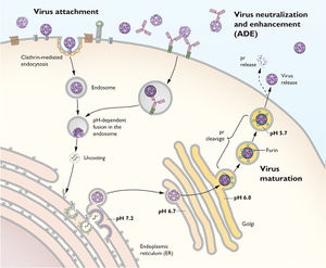
[Image:Flavivirus_Life_Cycle.jpg|thumb|300px|right|Diagram of the Flavivirus' life cycle. Notice the initial binding to the host cell and the clathrin mediated endocytosis that follows the initial binding. Then the viral membrane fuses with the host's endosome to expose the viral ssRNA to the endoplasmic reticulum (ER). There, the ssRNA is transcribed and copied to make new viral proteins and new ssRNA. The viral particles are assembled and form a immature virion by budding off of the ER. After budding off, the immature virion moves to the Golgi. Moving from the Golgi to the host's membrane, the cell undergoes cleavages that cleave the prM protein into M proteins, maturing the virion before being secreted out of the host cell. Additionally, notice how the immature virion has a rough, spiked surface in contrast to the smooth surface of the mature virion. Diagram by Ted C. Pierson, NIH National Institute of Allergy and Infectious Diseases]] | [[Image:Flavivirus_Life_Cycle.jpg|thumb|300px|right|Diagram of the Flavivirus' life cycle. Notice the initial binding to the host cell and the clathrin mediated endocytosis that follows the initial binding. Then the viral membrane fuses with the host's endosome to expose the viral ssRNA to the endoplasmic reticulum (ER). There, the ssRNA is transcribed and copied to make new viral proteins and new ssRNA. The viral particles are assembled and form a immature virion by budding off of the ER. After budding off, the immature virion moves to the Golgi. Moving from the Golgi to the host's membrane, the cell undergoes cleavages that cleave the prM protein into M proteins, maturing the virion before being secreted out of the host cell. Additionally, notice how the immature virion has a rough, spiked surface in contrast to the smooth surface of the mature virion. Diagram by Ted C. Pierson, NIH National Institute of Allergy and Infectious Diseases]] | ||
==Section 1== | ==Section 1== | ||
<br>Include some current research, with at least one figure showing data.<br> | <br>Include some current research, with at least one figure showing data.<br> | ||
Revision as of 05:12, 22 April 2014
Introduction
By Christopher Kei Helm
At right is a sample image insertion. It works for any image uploaded anywhere to MicrobeWiki. The insertion code consists of:
Double brackets: [[
Filename: PHIL_1181_lores.jpg
Thumbnail status: |thumb|
Pixel size: |300px|
Placement on page: |right|
Legend/credit: Electron micrograph of the Ebola Zaire virus. This was the first photo ever taken of the virus, on 10/13/1976. By Dr. F.A. Murphy, now at U.C. Davis, then at the CDC.
Closed double brackets: ]]
Other examples:
Bold
Italic
Subscript: H2O
Superscript: Fe3+
By Student Name
At right is a sample image insertion. It works for any image uploaded anywhere to MicrobeWiki. The insertion code consists of:
Double brackets: [[
Filename: PHIL_1181_lores.jpg
Thumbnail status: |thumb|
Pixel size: |300px|
Placement on page: |right|
Legend/credit: Electron micrograph of the Ebola Zaire virus. This was the first photo ever taken of the virus, on 10/13/1976. By Dr. F.A. Murphy, now at U.C. Davis, then at the CDC.
Closed double brackets: ]]
Other examples:
Bold
Italic
Subscript: H2O
Superscript: Fe3+
Introduce the topic of your paper. What microorganisms are of interest? Habitat? Applications for medicine and/or environment?

Section 1
Include some current research, with at least one figure showing data.


