Herpesviridae and Their Role in Uveitis: Difference between revisions
No edit summary |
No edit summary |
||
| Line 75: | Line 75: | ||
References | References | ||
1.↑ Hodgkin, J. and Partridge, F.A. "Caenorhabditis elegans meets microsporidia: the nematode killers from Paris." 2008. PLoS Biology 6:2634-2637. | 1.↑ Hodgkin, J. and Partridge, F.A. "Caenorhabditis elegans meets microsporidia: the nematode killers from Paris." 2008. PLoS Biology 6:2634-2637. | ||
2.↑ Bartlett et al.: Oncolytic viruses as therapeutic cancer vaccines. Molecular Cancer 2013 12:103. | 2.↑ Bartlett et al.: Oncolytic viruses as therapeutic cancer vaccines. Molecular Cancer 2013 12:103. | ||
3. Errera M.H., Goldschmidt, P., Batellier, L., Degorge S., Héron, E., Laroche, L., Sahel, J.A., Westcott, M., and Chaumeil, C."Real-time polymerase chain reaction and intraocular antibody production for the diagnosis of viral versus toxoplasmic infectious posterior uveitis." 2011. Graefe's Archive for Clinical and Experimental Ophthalmology. V 249 (12): 1837-1846. | |||
4. http://phil.cdc.gov/PHIL_Images/08301998/00014/B82-0474_lores.jpg | |||
Revision as of 03:22, 23 April 2016
Contents [hide] 1 Section 2 Section 1 3 Section 2 4 Section 3 5 Section 4 6 Conclusion 7 References
Section
(thumbnail)
Electron micrograph of the Ebola Zaire virus. This was the first photo ever taken of the virus, on 10/13/1976. By Dr. F.A. Murphy, now at U.C. Davis, then at the CDC.
By Chris Link
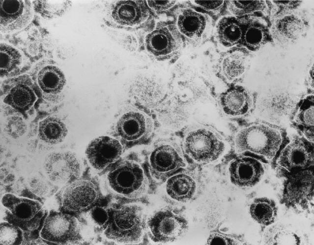
[[ Filename: Transmission_electron_micrograph_of_herpes_simplex_virus.jpg Thumbnail status: |thumb| Pixel size: |300px| Placement on page: |right| Legend/credit: Transmission electron micrograph of herpes simplex virus. Taken by Erskine Palmer - http://phil.cdc.gov/PHIL_Images/08301998/00014/B82-0474_lores.jpg ]]
At right is a sample image insertion. It works for any image uploaded anywhere to MicrobeWiki.
The insertion code consists of:
Double brackets: [[ Filename: PHIL_1181_lores.jpg Thumbnail status: |thumb| Pixel size: |300px| Placement on page: |right| Legend/credit: Electron micrograph of the Ebola Zaire virus. This was the first photo ever taken of the virus, on 10/13/1976. By Dr. F.A. Murphy, now at U.C. Davis, then at the CDC. Closed double brackets: ]]
Other examples:
Bold Italic Subscript: H2O Superscript: Fe3+
Introduce the topic of your paper. What is your research question? What experiments have addressed your question? Applications for medicine and/or environment?
Sample citations: [1] [2]
A citation code consists of a hyperlinked reference within "ref" begin and end codes.
Section 1
Include some current research, with at least one figure showing data.
Every point of information REQUIRES CITATION using the citation tool shown above.
Section 2
Include some current research, with at least one figure showing data.
Section 3
Include some current research, with at least one figure showing data.
Section 4
Conclusion
References 1.↑ Hodgkin, J. and Partridge, F.A. "Caenorhabditis elegans meets microsporidia: the nematode killers from Paris." 2008. PLoS Biology 6:2634-2637. 2.↑ Bartlett et al.: Oncolytic viruses as therapeutic cancer vaccines. Molecular Cancer 2013 12:103.
3. Errera M.H., Goldschmidt, P., Batellier, L., Degorge S., Héron, E., Laroche, L., Sahel, J.A., Westcott, M., and Chaumeil, C."Real-time polymerase chain reaction and intraocular antibody production for the diagnosis of viral versus toxoplasmic infectious posterior uveitis." 2011. Graefe's Archive for Clinical and Experimental Ophthalmology. V 249 (12): 1837-1846. 4. http://phil.cdc.gov/PHIL_Images/08301998/00014/B82-0474_lores.jpg
Authored for BIOL 238 Microbiology, taught by Joan Slonczewski, 2016, Kenyon College.
Category: ##Curated Pages
