Streptococcus pneumoniae: Difference between revisions
| Line 17: | Line 17: | ||
CDC Public Health Image Library. Public domain.]] | CDC Public Health Image Library. Public domain.]] | ||
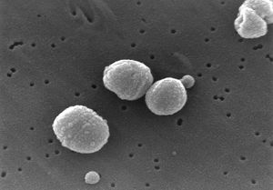
''Streptococcus pneumoniae'' are Gram-positive bacteria in the shape of a slightly pointed cocci. They are usually found in pairs (diplococci), but are also found singly and in short chains. ''S. pneumoniae'' are alpha hemolytic, a term describing how the cultured bacteria break down red blood cells for the purpose of classification. Individual bacteria are between 0.5 and 1.25 micrometers in diameter. ''S. pneumoniae'' do not form spores and are non-motile, though they sometimes have pili used for adherence. (5) | ''Streptococcus pneumoniae'' are Gram-positive bacteria in the shape of a slightly pointed cocci. They are usually found in pairs (diplococci), but are also found singly and in short chains. ''S. pneumoniae'' are alpha hemolytic, a term describing how the cultured bacteria break down red blood cells for the purpose of classification. Individual bacteria are between 0.5 and 1.25 micrometers in diameter. ''S. pneumoniae'' do not form spores and are non-motile, though they sometimes have pili used for adherence. (5) ''S. pneumonia'' are found normally in the upper respiratory tract, including the throat and nasal passages. (2) | ||
==Genome structure== | ==Genome structure== | ||
Revision as of 08:17, 6 June 2007
A Microbial Biorealm page on the genus Streptococcus pneumoniae
Classification
Higher order taxa
Bacteria; Firmicutes; Bacilli; Lactobacillales; Streptococcaceae
Species
Streptococcus pneumoniae
Description and significance
Streptococcus pneumoniae are Gram-positive bacteria in the shape of a slightly pointed cocci. They are usually found in pairs (diplococci), but are also found singly and in short chains. S. pneumoniae are alpha hemolytic, a term describing how the cultured bacteria break down red blood cells for the purpose of classification. Individual bacteria are between 0.5 and 1.25 micrometers in diameter. S. pneumoniae do not form spores and are non-motile, though they sometimes have pili used for adherence. (5) S. pneumonia are found normally in the upper respiratory tract, including the throat and nasal passages. (2)
Genome structure
The genome of "S. pneumoniae" consists of 2.16 million base pairs and contains 2236 predicted coding regions, 64% of which have been assigned a biological role. About 5% of the genome is made up of insertion sequences that may contribute to genome rearrangements through transferral of DNA. Several surface proteins have been identified that may serve as vaccine candidates. Several strains of "Streptococcus pneumoniae" have been identified, possibly accounting for differences in virulence and antigenicity.
Cell structure and metabolism
"Streptococcus pneumonia" gets a significant amount of its carbon and nitrogen through extracellular enzyme systems that allow the metabolism of polysaccharides and hexosamines, as well as cause damage to host tissue and enable colonization.
"Like other streptococci, they lack catalase and ferment glucose to lactic acid. Unlike other streptococci, they do not display an M protein, they hydrolyze inulin, and their cell wall composition is characteristic both in terms of their peptidoglycan and their teichoic acid)." (5)
Ecology
Pathology
Streptococcus pneumoniae is known to cause bacteremia, otitis media, and meningitis in humans, though it is best known for causing pneumonia, a disease of the upper respiratory tract that causes illness and death all over the world. (5) Symptoms of pneumonia include a cough accompanied by greenish or yellow mucous, fever, chills, shortness of breath, and chest pain. The bacteria enter the body most commonly via inhalation of small water droplets. Very young children and the elderly are the most prone to catching pneumonia.
The virulence factors of S. pneumoniae include a plysaccharide capsule that prevents phagocytosis by the host's immune cells, surface proteins that prevent the activation of complement (part of the immune system that helps clear pathogens from the body), and pili that enable S. pneumoniae to attach to epithelial cells in the upper respiratory tract. (4) Pili are long, thin extracellular organelles that are able to extend outside of the polysaccharide capsule. They are encoded by the rlrA islet (an area of a genome in which rapid mutation takes place) which is present in only some isolated strains of S. pneumoniae. These pili contribute to adherence and virulence, as well as increase the inflammatory response of the host. (4)
Disease caused by Streptococcus pneumoniae is most prevalent in poorer countries, increasing the need for an economical and effective vaccine. A potential candidate is the bacterium Lactococcus lactis, which produces the S. Pneumoniae surface protein A (PspA). Exposure to this strain may stimulate antibody production effective against S. Pneumoniae without running the risk of contracting the disease. (3)
Application to Biotechnology
Current Research
References
1. Hervé, T., et al. "Complete Genome Sequence of a Virulent Isolate of Streptococcus pneumoniae". Science. July 20, 2001. Volume 293. p. 498-506.
http://www.sciencemag.org/cgi/content/abstract/293/5529/498
2. Winstead, E. "Sugar Transporters and Foreign DNA, The sequenced Streptococcus pneumoniae genome". Genome News Network. July 23, 2001.
http://www.genomenewsnetwork.org/articles/07_01/Streptococcus_p.shtml
3. Hanniffy, S., Carter, A., Hitchin, E., and Wells, J. "Mucosal Delivery of a Pneumococcal Vaccine Using Lactococcus lactis Affords Protection against Respiratory Infection". The Journal of Infectious Diseases. 2007. Volume 195. p. 185-193.
4. Barocchi, M., Ries, J., Zogaj, X., Hemsley, C., Albiger, B., Kanth, A., Dahlberg, S., Fernebro, J., Moschioni, M., Masignani, V., Hultenby, K., Taddei, A., Beiter, K., Wartha, F., von Euler, A., Covacci, A., Holden, D., Normark, S., Rappuoli, R., Henriques-Normark, B. "A pneumococcal pilus influences virulence and host inflammatory responses". Proceedings of the National Academy of Sciences of the United States of America. 2006. Volume 103. p. 2857-62.
http://www.ncbi.nlm.nih.gov/sites/entrez?cmd=Retrieve&db=pubmed&dopt=Abstract&list_uids=16481624
5. Todar, K. "Streptococcus pneumoniae: Pneumococcal pneumonia". Todar's Online Textbook of Bacteriology. 2003.

