Acanthamoeba polyphaga: Difference between revisions
| Line 27: | Line 27: | ||
==Classification== | ==Classification== | ||
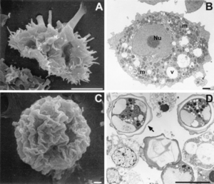
[[Image:classification 1.png|thumb|300px|right|(Left) Scanning (A and C) and transmission (B and D) electron micrographs depict the life cycle stages of Acanthamoeba spp. (A) A. polyphaga trophozoite; (B) trophozoite of A. castellanii showing the prominent central nucleolus (Nu), mitochondria (m), and cytoplasmid food vacuoles (v); (C) wrinkled cyst of A. polyphaga; and (D) double-walled cyst of A. cestellanii. Bars, 10 µm (A and D) and 1 µm (B and C). This photo was published in 2003. By Francine Marciano-Cabral and Guy Cabral [1].]] | [[Image:classification 1.png|thumb|300px|right|(Left) Scanning (A and C) and transmission (B and D) electron micrographs depict the life cycle stages of <i>Acanthamoeba</i> spp. (A) <i>A. polyphaga</i> trophozoite; (B) trophozoite of <i>A. castellanii</i> showing the prominent central nucleolus (Nu), mitochondria (m), and cytoplasmid food vacuoles (v); (C) wrinkled cyst of <i>A. polyphaga</i>; and (D) double-walled cyst of <i>A. cestellanii</i>. Bars, 10 µm (A and D) and 1 µm (B and C). This photo was published in 2003. By Francine Marciano-Cabral and Guy Cabral [1].]] | ||
Acanthamoeba are termed amphizoic organisms due to their ability to exist as either free-living or parasitic. Identification at the genus level is easy to detect since all Acanthamoeba trophozoites contain spiny projections on the surfaces of their cell [1]. | <i>Acanthamoeba</i> are termed amphizoic organisms due to their ability to exist as either free-living or parasitic. Identification at the genus level is easy to detect since all <i>Acanthamoeba</i> trophozoites contain spiny projections on the surfaces of their cell [1]. | ||
The species level has three morphological groups (I, II, and III) based on cyst size and shape. Species I have larger cysts, species II have a wrinkled outer cyst and a stellate inner cyst, and species III have a smooth ectocyst and a round endocyst. <i>A. polyphaga<i | The species level has three morphological groups (I, II, and III) based on cyst size and shape. Species I have larger cysts, species II have a wrinkled outer cyst and a stellate inner cyst, and species III have a smooth ectocyst and a round endocyst. <i>A. polyphaga</i> is placed in the Species II group since it is characterized as having a wrinkled ectocyst and a stellate endocyst [1]. | ||
[[Image:classification 2.png|thumb|300px|right|Phylogenetic tree showing Acanthamoeba at the genus level. This photo was published in 2003. By Francine Marciano-Cabral and Guy Cabral [1].]] | |||
==Section 2== | ==Section 2== | ||
Revision as of 03:11, 24 March 2013
Introduction

Acanthamoeba polyphaga is a free-living amoeba found in environments including soil, dust, air, seawater, tap water, and swimming pools [1].
A. polyphaga receives nutrition by consuming bacteria, algae, and yeast via phagocytosis. The amoeba ingests liquids through the process of pinocytosis. A. polyphaga acts as a secondary decomposer to remineralize soil with carbon, nitrogen, and phosphorous by consuming bacterial primary decomposers. Primary decomposers decompose organic material into minerals, but can not release these minerals from their own mass. After A. polyphaga digests the primary decomposers, the amoeba uses contractile vacuoles to release the environmentally useful bacterial nutrients from its food vacuole into the soil [1].
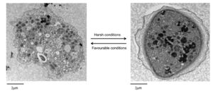
A. polyphaga, like the rest of the Acanthamoeba species, is divided into two lifecyles: the active trophozoite stage which reproduces via binary fission and the dormant, non-dividing cyst stage. Acanthamoeba polyphaga trophozoites contain one or more contractile vacuoles, lysosomes, digestive vacuoles, glycogen-containing vacuoles, a Golgi complex, smooth and rough endoplasmic reticulum, free ribosomes, microtubules, large numbers of mitochondria, and a single nucleus [1, 2]. The cysts of A. polyphaga contain durable double walls for protection in harsh conditions. The trophozoite turns into a cyst under adverse environmental conditions such as during food deprivation, desiccation, and changes in temperature and pH [1].
Other examples:
Bold
Italic
Subscript: H2O
Superscript: Fe3+
Classification

Acanthamoeba are termed amphizoic organisms due to their ability to exist as either free-living or parasitic. Identification at the genus level is easy to detect since all Acanthamoeba trophozoites contain spiny projections on the surfaces of their cell [1].
The species level has three morphological groups (I, II, and III) based on cyst size and shape. Species I have larger cysts, species II have a wrinkled outer cyst and a stellate inner cyst, and species III have a smooth ectocyst and a round endocyst. A. polyphaga is placed in the Species II group since it is characterized as having a wrinkled ectocyst and a stellate endocyst [1].
Section 2
Include some current research in each topic, with at least one figure showing data.
Section 3
Include some current research in each topic, with at least one figure showing data.
Conclusion
Overall paper length should be 3,000 words, with at least 3 figures.
References
Edited by (your name here), a student of Nora Sullivan in BIOL187S (Microbial Life) in The Keck Science Department of the Claremont Colleges Spring 2013.
