Bacillus Anthracis: Anthrax Lethal Toxin: Difference between revisions
No edit summary |
No edit summary |
||
| Line 15: | Line 15: | ||
<br><b>Superscript:</b> Fe<sup>3+</sup> | <br><b>Superscript:</b> Fe<sup>3+</sup> | ||
[[Image:Bacillus_anthracis_gram.jpg|thumb|300px| | <p>Anthrax is a disease that typically affects herbivores but can infect any mammal, including humans. It is caused by the bacterium Bacillus anthracis (B. anthracis). This disease has become a hot topic in today’s society, in which terrorism is becoming more prevalent, as it can be used in biological warfare. Recently, there has also been an increase in the number of cases of injection anthrax, a form of the disease that affects heroin users, in Europe (Grunow). However, this disease is not new. In fact, there is evidence of it throughout history. The fifth and sixth plagues in Egypt (Exodus, Chapter 9) have been attributed to this pathogen (Baillie). </p> | ||
<p>The species derives its name from the Greek word anthrakis, meaning coal, because infection by the organism causes the formation of black, cutaneous eschars (Spencer). There are are three types of B. anthracis infection that are typically described: cutaneous anthrax (the cause of the black sores), inhalation anthrax (the most deadly form), and gastrointestinal anthrax (Mock, Mignot).</p> | |||
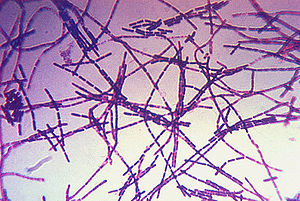
[[Image:Bacillus_anthracis_gram.jpg|thumb|300px|right|A photomicrograph of Bacillus anthracis bacteria using Gram-stain technique. Photo obtained from the CDC's Public Health Image Library.]] | |||
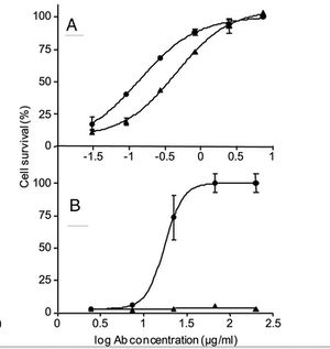
[[Image:cAb29Fig.jpg|thumb|300px|center|Figure 1. A and B show the neutralization assay performed using cultured J774A macrophages that were incubated for 5 h with fixed amounts of LF and PA83 (A) or purified prepore (B) in the presence of cAb29 (circles) or Ab33 (triangles) in the indicated concentrations. Cell survival was determined by XTT and plotted as the percentage of untreated control cells. Points are mean ± S.D. of triplicate determinants. Ab concentration, antibody concentration (Mechaly).]] | [[Image:cAb29Fig.jpg|thumb|300px|center|Figure 1. A and B show the neutralization assay performed using cultured J774A macrophages that were incubated for 5 h with fixed amounts of LF and PA83 (A) or purified prepore (B) in the presence of cAb29 (circles) or Ab33 (triangles) in the indicated concentrations. Cell survival was determined by XTT and plotted as the percentage of untreated control cells. Points are mean ± S.D. of triplicate determinants. Ab concentration, antibody concentration (Mechaly).]] | ||
| Line 27: | Line 31: | ||
<br>Introduce the topic of your paper. What microorganisms are of interest? Habitat? Applications for medicine and/or environment?<br> | <br>Introduce the topic of your paper. What microorganisms are of interest? Habitat? Applications for medicine and/or environment?<br> | ||
== | ==Organism== | ||
<br>Include some current research, with at least one figure showing data.<br> | <br>Include some current research, with at least one figure showing data.<br> | ||
== | ==Life Cycle== | ||
<br>Include some current research, with at least one figure showing data.<br> | <br>Include some current research, with at least one figure showing data.<br> | ||
== | ==Pathogenesis== | ||
<br>Include some current research, with at least one figure showing data.<br> | <br>Include some current research, with at least one figure showing data.<br> | ||
== | ==Clinical Symptoms and Diagnosis== | ||
<br>Overall text length at least 3,000 words, with at least 3 figures.<br> | <br>Overall text length at least 3,000 words, with at least 3 figures.<br> | ||
==Anthrax Toxin Neutralization with Antibody== | |||
==Target Cells in Humans in Inhalation Anthrax== | |||
==References== | ==References== | ||
Revision as of 02:23, 25 April 2013
Introduction
By Connor Gibbons
At right is a sample image insertion. It works for any image uploaded anywhere to MicrobeWiki. The insertion code consists of:
Double brackets: [[
Filename: PHIL_1181_lores.jpg
Thumbnail status: |thumb|
Pixel size: |300px|
Placement on page: |right|
Legend/credit: Electron micrograph of the Ebola Zaire virus. This was the first photo ever taken of the virus, on 10/13/1976. By Dr. F.A. Murphy, now at U.C. Davis, then at the CDC.
Closed double brackets: ]]
Other examples:
Bold
Italic
Subscript: H2O
Superscript: Fe3+
Anthrax is a disease that typically affects herbivores but can infect any mammal, including humans. It is caused by the bacterium Bacillus anthracis (B. anthracis). This disease has become a hot topic in today’s society, in which terrorism is becoming more prevalent, as it can be used in biological warfare. Recently, there has also been an increase in the number of cases of injection anthrax, a form of the disease that affects heroin users, in Europe (Grunow). However, this disease is not new. In fact, there is evidence of it throughout history. The fifth and sixth plagues in Egypt (Exodus, Chapter 9) have been attributed to this pathogen (Baillie).
The species derives its name from the Greek word anthrakis, meaning coal, because infection by the organism causes the formation of black, cutaneous eschars (Spencer). There are are three types of B. anthracis infection that are typically described: cutaneous anthrax (the cause of the black sores), inhalation anthrax (the most deadly form), and gastrointestinal anthrax (Mock, Mignot).


Introduce the topic of your paper. What microorganisms are of interest? Habitat? Applications for medicine and/or environment?
Organism
Include some current research, with at least one figure showing data.
Life Cycle
Include some current research, with at least one figure showing data.
Pathogenesis
Include some current research, with at least one figure showing data.
Clinical Symptoms and Diagnosis
Overall text length at least 3,000 words, with at least 3 figures.
Anthrax Toxin Neutralization with Antibody
Target Cells in Humans in Inhalation Anthrax
References
Edited by student of Joan Slonczewski for BIOL 238 Microbiology, 2011, Kenyon College.

