Magnetotactic Bacteria: Difference between revisions
No edit summary |
No edit summary |
||
| Line 4: | Line 4: | ||
[[Image:magnetosomeformation.jpeg|thumb|300px|right| <b>Figure 3</b>- Proposed model of magnetosome formation. "First, the reshaping of the inner cell membrane by MamI, MamL, MamQ, MamB, and other factors (green) creates the magnetosome membrane. Second, MamE is recruited to the nascent magnetosome. Third, MamE (in blue), independent of its protease activity recruits other proteins (red) to the magnetosome. Fourth, MamK and MamJ help to organize magnetosomes into chains. Note that this step is independent of biomineralization and may occur before or after crystal formation. Fifth, biomineralization is initiated, and small crystals of magnetite are formed. Finally, these small crystals are matured into large crystals in a step that requires the proteolytic activity of MamE." Komeili (2012) Courtesy of Komeili (http://femsre.oxfordjournals.org/content/36/1/232.figures-only)]] | [[Image:magnetosomeformation.jpeg|thumb|300px|right| <b>Figure 3</b>- Proposed model of magnetosome formation. "First, the reshaping of the inner cell membrane by MamI, MamL, MamQ, MamB, and other factors (green) creates the magnetosome membrane. Second, MamE is recruited to the nascent magnetosome. Third, MamE (in blue), independent of its protease activity recruits other proteins (red) to the magnetosome. Fourth, MamK and MamJ help to organize magnetosomes into chains. Note that this step is independent of biomineralization and may occur before or after crystal formation. Fifth, biomineralization is initiated, and small crystals of magnetite are formed. Finally, these small crystals are matured into large crystals in a step that requires the proteolytic activity of MamE." Komeili (2012) Courtesy of Komeili (http://femsre.oxfordjournals.org/content/36/1/232.figures-only)]] | ||
<br> | <br> | ||
Magnetotactic bacteria (MB) are [http://faculty.ccbcmd.edu/courses/bio141/lecguide/unit1/prostruct/gncw.html gram-negative] bacteria that build specialized organelles called [http://www.ncbi.nlm.nih.gov/pubmed/23615196 magnetosomes] in order to store magnetic material and align themselves with the earth’s magnetic field. Magnetotactic bacteria were first described in 1975 when Richard Blakemore realized that a specific group of bacteria he collected from sediment constantly swam in the same geographic direction, regardless of the positioning of the microscope or external stimuli < | Magnetotactic bacteria (MB) are [http://faculty.ccbcmd.edu/courses/bio141/lecguide/unit1/prostruct/gncw.html gram-negative] bacteria that build specialized organelles called [http://www.ncbi.nlm.nih.gov/pubmed/23615196 magnetosomes] in order to store magnetic material and align themselves with the earth’s magnetic field. Magnetotactic bacteria were first described in 1975 when Richard Blakemore realized that a specific group of bacteria he collected from sediment constantly swam in the same geographic direction, regardless of the positioning of the microscope or external stimuli <ref>[http://www.sciencemag.org/content/190/4212/377.short Blakemore, R. (1975). Magnetotactic bacteria. Science (New York, N.Y.), 190(4212), 377-379.]</ref>. MB are mostly found in shallow aquatic environments where oxygen and other redox compounds are horizontally stratified and many described magnetotactic bacteria localize at or close to the oxic anoxic transition zone (OATZ)—a region in the water column that has very low oxygen levels <sup>[2]</sup>. The current model (shown in Figure 1) to explain the selective advantage provided by magnetosomes is that magnetotactic bacteria are able to locate the OATZ much easier than bacteria that solely use [http://en.wikipedia.org/wiki/Chemotaxis chemotactic] and [http://medical-dictionary.thefreedictionary.com/aerotaxis aerotactic] mechanisms <sup>[3]</sup>. Although the magneto-aerotaxis model has been widely accepted amongst the scientific community, new research is suggesting that the behavior magnetotactic bacteria exhibit in the environment may be more complicated than a simple response to oxygen levels: | ||
<br> | <br> | ||
<br> | <br> | ||
| Line 51: | Line 51: | ||
[10] [http://link.springer.com/article/10.1007/BF00169632 Matsunaga, T., Sakaguchi, T., & Tadakoro, F. (1991). Magnetite formation by a magnetic bacterium capable of growing aerobically. Applied Microbiology and Biotechnology, 35(5), 651-655.] | [10] [http://link.springer.com/article/10.1007/BF00169632 Matsunaga, T., Sakaguchi, T., & Tadakoro, F. (1991). Magnetite formation by a magnetic bacterium capable of growing aerobically. Applied Microbiology and Biotechnology, 35(5), 651-655.] | ||
[11] [http://www.sciencedirect.com/science/article/pii/S0723202011803139 Schleifer, K. H., Schüler, D., Spring, S., Weizenegger, M., Amann, R., Ludwig, W., et al. (1991). The genus magnetospirillum gen. nov. description of magnetospirillum gryphiswaldense sp. nov. and transfer of aquaspirillum magnetotacticum to magnetospirillum magnetotacticum comb. nov. Systematic and Applied Microbiology, 14(4), 379-385.] | [11] [http://www.sciencedirect.com/science/article/pii/S0723202011803139 Schleifer, K. H., Schüler, D., Spring, S., Weizenegger, M., Amann, R., Ludwig, W., et al. (1991). The genus magnetospirillum gen. nov. description of magnetospirillum gryphiswaldense sp. nov. and transfer of aquaspirillum magnetotacticum to magnetospirillum magnetotacticum comb. nov. Systematic and Applied Microbiology, 14(4), 379-385.] | ||
| Line 59: | Line 58: | ||
[] [http://www.sciencedirect.com/science/article/pii/S0065216407620024 Bazylinski, D. A., & Schübbe, S. (2007). Controlled biomineralization by and applications of magnetotactic bacteria. Advances in applied microbiology, 62, 21-62.] | [] [http://www.sciencedirect.com/science/article/pii/S0065216407620024 Bazylinski, D. A., & Schübbe, S. (2007). Controlled biomineralization by and applications of magnetotactic bacteria. Advances in applied microbiology, 62, 21-62.] | ||
References: | |||
{{reflist}} | |||
<!--Do not edit or remove this line-->[[Category:Pages edited by students of Joan Slonczewski at Kenyon College]] | <!--Do not edit or remove this line-->[[Category:Pages edited by students of Joan Slonczewski at Kenyon College]] | ||
Revision as of 07:51, 24 March 2015
Introduction to Magnetotactic Bacteria


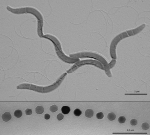
Magnetotactic bacteria (MB) are gram-negative bacteria that build specialized organelles called magnetosomes in order to store magnetic material and align themselves with the earth’s magnetic field. Magnetotactic bacteria were first described in 1975 when Richard Blakemore realized that a specific group of bacteria he collected from sediment constantly swam in the same geographic direction, regardless of the positioning of the microscope or external stimuli [1]. MB are mostly found in shallow aquatic environments where oxygen and other redox compounds are horizontally stratified and many described magnetotactic bacteria localize at or close to the oxic anoxic transition zone (OATZ)—a region in the water column that has very low oxygen levels [2]. The current model (shown in Figure 1) to explain the selective advantage provided by magnetosomes is that magnetotactic bacteria are able to locate the OATZ much easier than bacteria that solely use chemotactic and aerotactic mechanisms [3]. Although the magneto-aerotaxis model has been widely accepted amongst the scientific community, new research is suggesting that the behavior magnetotactic bacteria exhibit in the environment may be more complicated than a simple response to oxygen levels:
- Some MB species also show phototactic response, which helps reinforce magneto-aerotactic behavior and repel them from surface waters [4, 5]
- Genome sequences show that MB have some of the highest numbers of signaling proteins of Bacteria [6]
- MB produce more magnetosomes than necessary to align with the earth’s magnetic field [7]
- MB have been found near the equator, where their magneto-aerotactic behavior has no advantage [8]
Regardless of the actual biological function of magnetosomes, magnetotactic bacteria are a very interesting research topic because they have the potential to impact a large number of scientific and applied disciplines. Magnetosomes are the perfect model for the study of cellular compartmentalization and organization in bacteria because the formation of specialized, membrane bound, features is usually attributed to eukaryotic organisms. Magnetotactic bacteria are also a great model for the study of biomineralization because of the precise control over the composition, size and morphology of magenetite crystals in magnetosomes.
Magnetosome Island
Bacterial genomes can evolve over time due to mutations, rearrangements, or horizontal gene transfer and many genes that are acquired through horizontal gene transfer come in blocks that are recognized as genomic islands. These genomic islands can be recognized by nucleotide statistics (GC skew) that differ from the rest of the organisms DNA and are often associated with inserted tRNA genes, pseudogenes, transposons, and IS elements[9].
Magnetosome Formation
Identification of the genetic elements needed for magnetosome formation took many years due the lack of cultured and genetically tractable model organisms. Aquaspirillum magnetotacticum, now referred to as Magnetospirillum magnetotacticum, or MS-1, was the first magnetotactic bacteria to be isolated in pure culture. Two closely related Magnetospirillum species, Magnetospirillum grysphiswaldense (MSR-1) and Magnetospirillum magneticum (AMB-1), were isolated soon after and have become the focus of most magnetosome formation research[10,11]. Three approaches have been used to determine the molecular factors involved in the formation of magnetosomes: proteomics, genetic analysis, and comparative genomics.
Applications in Bioremediation
References
[1] Blakemore, R. (1975). Magnetotactic bacteria. Science (New York, N.Y.), 190(4212), 377-379.
Other References:
References:
Chockalingam, Evvie, and S. Subramanian. “Utility of Eucalyptus Tereticornis (Smith) Bark and Desulfotomaculum Nigrificans for the Remediation of Acid Mine Drainage.” Bioresource Technology 100, no. 2 (January 2009): 615–621. doi:10.1016/j.biortech.2008.07.004.
“Genus Desulfotomaculum - Hierarchy - The Taxonomicon.” Accessed November 5, 2013. http://taxonomicon.taxonomy.nl/TaxonTree.aspx?id=229.
Kaksonen, Anna H., Stefan Spring, Peter Schumann, Reiner M. Kroppenstedt, and Jaakko A. Puhakka. “Desulfotomaculum Thermosubterraneum Sp. Nov., a Thermophilic Sulfate-reducer Isolated from an Underground Mine Located in a Geothermally Active Area.” International Journal of Systematic and Evolutionary Microbiology 56, no. 11 (November 1, 2006): 2603–2608. doi:10.1099/ijs.0.64439-0.
Liu, Yitai, Tim M. Karnauchow, Ken F. Jarrell, David L. Balkwill, Gwendolyn R. Drake, David Ringelberg, Ronald Clarno, and David R. Boone. “Description of Two New Thermophilic Desulfotomaculum Spp., Desulfotomaculum Putei Sp. Nov., from a Deep Terrestrial Subsurface, and Desulfotomaculum Luciae Sp. Nov., from a Hot Spring.” International Journal of Systematic Bacteriology 47, no. 3 (July 1, 1997): 615–621. doi:10.1099/00207713-47-3-615.
Moser, Duane P, Thomas M Gihring, Fred J Brockman, James K Fredrickson, David L Balkwill, Michael E Dollhopf, Barbara Sherwood Lollar, et al. “Desulfotomaculum and Methanobacterium Spp. Dominate a 4- to 5-kilometer-deep Fault.” Applied and Environmental Microbiology 71, no. 12 (December 2005): 8773–8783. doi:10.1128/AEM.71.12.8773-8783.2005.
Ogg, Christopher D, and Bharat K C Patel. “Desulfotomaculum Varum Sp. Nov., a Moderately Thermophilic Sulfate-reducing Bacterium Isolated from a Microbial Mat Colonizing a Great Artesian Basin Bore Well Runoff Channel.” 3 Biotech 1, no. 3 (October 2011): 139–149. doi:10.1007/s13205-011-0017-5.
Pikuta, E, A Lysenko, N Suzina, G Osipov, B Kuznetsov, T Tourova, V Akimenko, and K Laurinavichius. “Desulfotomaculum Alkaliphilum Sp. Nov., a New Alkaliphilic, Moderately Thermophilic, Sulfate-reducing Bacterium.” International Journal of Systematic and Evolutionary Microbiology 50 Pt 1 (January 2000): 25–33.
