THE DIAGNOSIS OF SYPHILIS: Difference between revisions
| Line 8: | Line 8: | ||
==Section 1== | ==Section 1== | ||
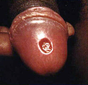
Before understand how to diagnose syphilis, it is important to know about the stages of disease since different methods may be used for different stages. Primary syphilis h occurs 9-90 days after contact with <br><i>T. pallidum</i> (Tramont, 2005). It is manifested by a skin lesion called a chancre Figure 1) (CDC, 2013). It starts out as flat area of redness that develops into a bump and then into a swallowing opening in the skin. The chancre occurs at any place on the body in which an individual has had contact with T. pallidum. This is usually on the penis in a man or external genitalia in a woman but can be in the mouth or even on a finger. | Before understand how to diagnose syphilis, it is important to know about the stages of disease since different methods may be used for different stages. Primary syphilis h occurs 9-90 days after contact with <br><i>T. pallidum</i> (Tramont, 2005). It is manifested by a skin lesion called a chancre Figure 1) (CDC, 2013). It starts out as flat area of redness that develops into a bump and then into a swallowing opening in the skin. The chancre occurs at any place on the body in which an individual has had contact with T. pallidum. This is usually on the penis in a man or external genitalia in a woman but can be in the mouth or even on a finger. | ||
[[Image:chancre-penile-1.jpg|thumb|300px|right|Syphilis penis. Primary stage of syphilis (chancre) on glans (head) of the penis.[http://www.cdc.gov/std/syphilis/images/chancre-penile.htm].]] | |||
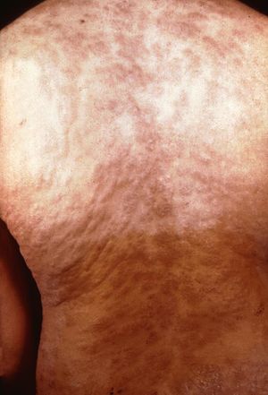
[[Image:Second stage syphilis.jpg |thumb|300px|right|Syphilis penis. Secondary stage of syphilis Rash on back.[http://www.cdc.gov/std/syphilis/images/rash-gbr.htm].]] | |||
The next stage is secondary syphilis. This occurs 3-10 weeks after the initial lesion if a person is not treated with antibiotics (Tramont, 2005). This means that T. pallidum has spread throughout the body. The most common symptom is a non-itchy rash that is not raised and often is on all parts of the body (Figure 2). Other manifestations can include fever, headache, weight loss, lymph node swelling, loss of hair, and eye disease. Individuals also may develop condyloma latum which are flat bumps in the genital area which have lots of T. pallidum and are very infectious. In addition, individuals can get flat patches in their mouth called mucous patches. | The next stage is secondary syphilis. This occurs 3-10 weeks after the initial lesion if a person is not treated with antibiotics (Tramont, 2005). This means that T. pallidum has spread throughout the body. The most common symptom is a non-itchy rash that is not raised and often is on all parts of the body (Figure 2). Other manifestations can include fever, headache, weight loss, lymph node swelling, loss of hair, and eye disease. Individuals also may develop condyloma latum which are flat bumps in the genital area which have lots of T. pallidum and are very infectious. In addition, individuals can get flat patches in their mouth called mucous patches. | ||
| Line 18: | Line 19: | ||
<br> | <br> | ||
<br> | <br> | ||
==Section 2== | ==Section 2== | ||
Revision as of 16:26, 21 April 2015
Introduction
By [Heather Fantry]
Syphilis is a sexually transmitted infection that has been rising in prevalence since 2000 (CDC, 2013). It is caused by the spirochete
Treponema pallidum.
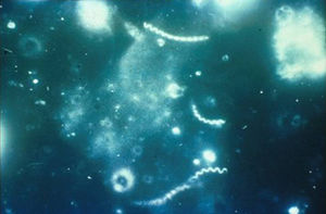
T. pallidum is a thin, tightly coiled spirochete that is microaerophilic (Tramont, 2005). Unlike most bacteria that infect humans, it cannot be cultured in the laboratory. It can only be cultured in laboratory animals, usually rabbits, which are not readily available in hospitals or medical clinics. Hence the diagnosis of syphilis is extremely difficult.
Bold
Italic
Section 1
Before understand how to diagnose syphilis, it is important to know about the stages of disease since different methods may be used for different stages. Primary syphilis h occurs 9-90 days after contact with
T. pallidum (Tramont, 2005). It is manifested by a skin lesion called a chancre Figure 1) (CDC, 2013). It starts out as flat area of redness that develops into a bump and then into a swallowing opening in the skin. The chancre occurs at any place on the body in which an individual has had contact with T. pallidum. This is usually on the penis in a man or external genitalia in a woman but can be in the mouth or even on a finger.


The next stage is secondary syphilis. This occurs 3-10 weeks after the initial lesion if a person is not treated with antibiotics (Tramont, 2005). This means that T. pallidum has spread throughout the body. The most common symptom is a non-itchy rash that is not raised and often is on all parts of the body (Figure 2). Other manifestations can include fever, headache, weight loss, lymph node swelling, loss of hair, and eye disease. Individuals also may develop condyloma latum which are flat bumps in the genital area which have lots of T. pallidum and are very infectious. In addition, individuals can get flat patches in their mouth called mucous patches.
The third stage is latent syphilis (CDC, 2013). It has no signs or symptoms and the only manifestation of the disease is a positive blood test.
The final stage is tertiary syphilis which only occurs in 35% of untreated patients (Tramont 2005). It occurs 10-25 years after primary syphilis. Most commonly it causes disease of the nervous system but it also can cause heart disease or deposits in the skin, soft tissue, or bones called gummas. The neurological manifestations are meningitis, strokes, dysfunction of cranial nerves, dementia, difficulty walking, visual or auditory problems, and loss of vibration sense.
T. pallidum can be transmitted from a mother to her unborn child which is called congenital syphilis (CDC, 2013). Although rare in the United States, it is of great concern because it can cause deafness, neurological problems, bone deformities, and even death (CDC, 2013).
Section 2
Include some current research, with at least one figure showing data.
Section 3
Include some current research, with at least one figure showing data.

References
[1] Hodgkin, J. and Partridge, F.A. "Caenorhabditis elegans meets microsporidia: the nematode killers from Paris." 2008. PLoS Biology 6:2634-2637.
Authored for BIOL 238 Microbiology, taught by Joan Slonczewski, 2015, Kenyon College.
