|
|
| Line 5: |
Line 5: |
|
| |
|
| <br>By Nadia Torok<br><br> | | <br>By Nadia Torok<br><br> |
| <i>Baylisascaris procyonis</i> is a parasitic nematode, or helminth, endemic to raccoon (<i>Procyon lotor</i>) <ref name=Page>[http://www.bioone.org/doi/pdf/10.1674/0003-0031%281998%29140%5B0180%3ARLSAIP%5D2.0.CO%3B2 Page, K.L., Swihart, R.K., and Kazacos, K.R. "Raccoon Latrine Structure and Its Potential Role in Transmission of <i>Baylisascaris procyonis</i> to vertebrates." 1998. The American Midland Naturalist 140(1):180-185.]</ref> populations<ref name=Gavin>[[https://www.ncbi.nlm.nih.gov/pmc/articles/PMC1265913/pdf/0058-04.pdf] Gavin, P.J., Kazacos, K.R., and Shulman, S.T. "Baylisascariasis." 2005. Clinical Microbiology Reviews 18(4): 703-718. DOI: 10.1128/CMR.18.4.703-718.2005]</ref>. This parasite causes baylisascariass in humans upon infection<ref name=Gavin/> and can infect other small mammals as well<ref name=Page/>. The species is distinguished from other helminthes by its invasion of the central nervous system (CNS) in hosts, as well as the continued growth of larvae within the CNS to a large size<ref name=Gavin/>. The first documented cases occurred in two young boys in 1984 and 1985<ref name=Gavin/>.
| | This is where my introductory paragraph will go. A citation example is posted below. <br> |
| <br><br>
| | |
| This parasite is responsible for various diseases in humans and other animals, which can be serious and often fatal in humans despite treatment<ref name=Gavin/>. Though infection with <i>B. procyonis</i> is rare and there have been fewer than 25 human cases reported in the United States<ref name=CDC>[[https://www.cdc.gov/parasites/baylisascaris/epi.html] "Parasites - Baylisascaris Infection." 2012. Centers for Disease Control Global Health - Division of Parasitic Diseases.]</ref>, infection is thought to be frequently misdiagnosed <ref name=CDC/> and prognosis is very poor<ref name=Gavin/>. The major clinical manifestations of <i>B. procyonis</i> include visceral larva migrans, neural larva migrans, and ocular larva migrans, which fall under the broader description of infection with <i>B. procyonis</i>, termed Baylisascaris<ref name=Gavin/>. | |
| <br><br>
| |
| <i>Baylisascaris procyonis</i> is a member of large nematodes in the order ascaridida, related to species such as <i>Ascaris lumbricoides, Toxocara canis</i>, and <i>Toxocara cati</i>, which are more commonly observed parasites affecting humans, dogs, or cats<ref name=Gavin/>. Originally, <i>B. procyonis </i>was classified as <i>Ascaris columnaris</i> in 1931, and it is closely related to <i>B. columnaris</i> and <i>B. melis</i>, other worms that parasitize skunks and badgers<ref name=Gavin/>.
| |
| <br><br>
| |
| These roundworms are tan in color and display sexual dimorphism, as females range in size from 20-22 cm and males from 9-11 cm <ref name=Gavin/>, making them some of the larger parasitic nematodes<ref name=Gavin/>. Females lay 115,000 to 179,000 eggs per day<ref name=Gavin/>. Eggs are an ellipsoidal shape, dark brown, and 63-88 μm by 50-70 μm in size <ref name=Gavin/>. It is estimated that raccoons shed up to 20,000 eggs per gram of feces<ref name=Page/>, while only 5,000 eggs or fewer is sufficient to infect a human <ref name=Nevada>[[http://southernnevadahealthdistrict.org/health-topics/baylisascaris-proyonis.php] Southern Nevada Health District. "Fact Sheet:
| |
| Raccoon Roundworm (Baylisascaris procyonis)." 2007. Southern Nevada Health District, Office of Epidemiology.]</ref>. An image of adult <i>B. procyonis</i> obtained from a raccoon can be seen at the right. <br><br>
| |
|
| |
|
| The insertion code consists of: | | The insertion code consists of: |
Revision as of 20:29, 12 April 2017
Introduction and Phylogeny
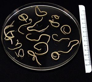
This is an image of Baylisascaris procyonis adults obtained from a raccoon. Image from
CDC.gov.
By Nadia Torok
This is where my introductory paragraph will go. A citation example is posted below.
The insertion code consists of:
Double brackets: [[
Filename: PHIL_1181_lores.jpg
Thumbnail status: |thumb|
Pixel size: |300px|
Placement on page: |right|
Legend/credit: Electron micrograph of the Ebola Zaire virus. This was the first photo ever taken of the virus, on 10/13/1976. By Dr. F.A. Murphy, now at U.C. Davis, then at the CDC.
Closed double brackets: ]]
Other examples:
Bold
Italic
Subscript: H2O
Superscript: Fe3+
Introduce the topic of your paper. What is your research question? What experiments have addressed your question? Applications for medicine and/or environment?
Sample citations: [1]
[2]
A citation code consists of a hyperlinked reference within "ref" begin and end codes.
Latrine habits: [3]
This is a test, according to a source.[3]
Section 1
Include some current research, with at least one figure showing data.
Every point of information REQUIRES CITATION using the citation tool shown above.
Section 2
Include some current research, with at least one figure showing data.
Section 3
Include some current research, with at least one figure showing data.
Section 4
Conclusion
References
Authored for BIOL 238 Microbiology, taught by Joan Slonczewski, 2017, Kenyon College.

