Vorticella: Difference between revisions
(Added an image.) |
(Added another image.) |
||
| Line 3: | Line 3: | ||
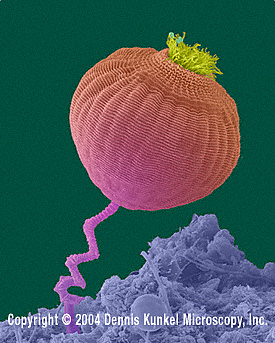
[[Image:99112C.jpg|frame|right|''Vorticella ''spp. [http://www.denniskunkel.com/index.php Dennis Kunkel Microscopy Inc].]] | [[Image:99112C.jpg|frame|right|''Vorticella ''spp. [http://www.denniskunkel.com/index.php Dennis Kunkel Microscopy Inc].]] | ||
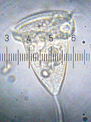
[[Image:20090920_213358_Vorticella.jpg|thumb|300px|right|A <i>Vorticella.</i> | [[Image:20090920_213358_Vorticella.jpg|thumb|300px|right|A <i>Vorticella.</i> | ||
<br/>Numbered ticks are 20 µM apart.<br/>Photograph by [[User:Blaylock|Bob Blaylock]]]] | <br/>Numbered ticks are 20 µM apart.<br/>Photograph by [[User:Blaylock|Bob Blaylock]].]] | ||
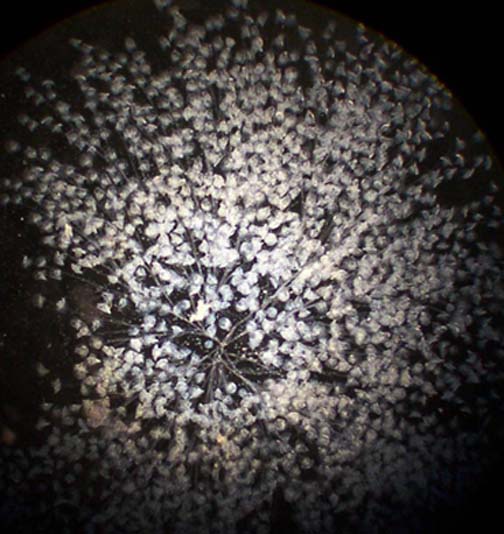
[[Image:20090402_0234_Vorticellas.jpg|thumb|300px|right|A bunch of <i>Vorticella.</i> | |||
<br/>Numbered ticks are 122 µM apart.<br/>Photograph by [[User:Blaylock|Bob Blaylock]].]] | |||
{| width="800" cellspacing="2" cellpadding="2" align="center" | {| width="800" cellspacing="2" cellpadding="2" align="center" | ||
Revision as of 08:02, 24 July 2010
A Microbial Biorealm page on the genus Vorticella

ClassificationHigher order taxa:Eukaryota; Alveolata; Ciliophora; Oligohymenophorea; Peritrichia; Vorticellidae.Species:Vorticella campanulaVorticella convallaria Vorticella microstoma
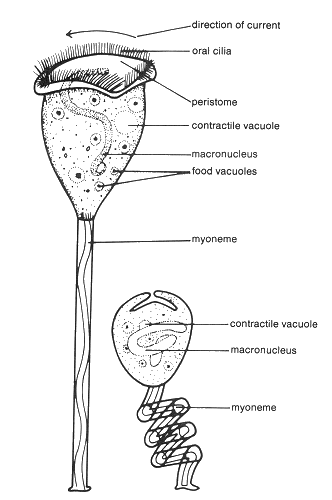
Description and SignificanceVorticella are members of the phylum Ciliophora. In some ways, they resemble members of the phylum Suctoria. However, there are major morphological differences between these two types of organisms. It is the unique structure of Vorticella that distinguishes them from other ciliates. Genome StructureLike some other ciliates, Vorticella has a deviant genetic code. UAA, a traditional stop codon, instead translates for glutamate. The small subunit rRNA (SSrRNA) gene has proved crucial for distinguishing between Vorticella species. Because different species are physically very similar, it is difficult to tell them apart by morphological characterstics alone. SSrRNA has proved a much more effective method of classification and identification. Cell Structure and Metabolism Diagram of Vorticella. Anabaena"Vorticella" lesson by the Haw River Program.
EcologyVorticella are aquatic organisms, most commonly found in freshwater habitats. They attach themselves to plant detritus, rocks, algae, or animals (particularly crustaceans). They are individual organisms, but often can be found in colonies. However, these are not true colonies, because each individual retains its own stalk. Vorticella are therefore free to separate from the colony at any time. A Vorticella colony. Photo by Howard Webb. References.Updated June 24, 2005 Haw River Program. "Vorticella." Accessed 23 June 2005. MicroscopeWorld.com. "Vorticella". 2005. Accessed 23 June 2005. |


