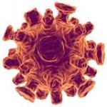
Cytomegalovirus in post transplant patients
Introduction

The cytomegalovirus (CMV) is a double stranded DNA virus part of the Herpesviridae viral family also known as herpesviruses. Herpesviruses share common characteristics including the ability to remain metabolically inert within the human body for extended periods of time. The human cytomegalovirus (HCMV) also known as Human herpesvirus 5 (HHV-5) is a species that causes infections typically associated with the salivary gland. HCMV is a common virus where about 50%-80% of the general adult population is infected (Slonczewski, 2013). However, most people who are infected are unaware since the virus is typically dormant in healthy individuals. The high prevalence of the virus among the general population is due to the many avenues of viral transmission. It is easily transmitted by person-to-person contact by saliva, urine, semen, blood, and cervical secretions during birth. Those with compromised immune systems manifest symptoms similar to mononucleosis (Epstein-Barr virus), and are affected in just about every organ in the body. Immunocompromised individuals such as organ transplant recipients and AIDS patients are especially at risk for more severe primary infections including latent infection, asymptomatic viral shedding (when the virus is active but doesn’t show visible signs or symptoms), and life-threatening multisystem disease (Farrugia, 1992). In such patients, HCMV not only causes problems in the acute phase but also increases the risk of long-term complications (Soderberg-Naucler, 2008).
CMV virology
Among the hundreds of herpes virus, there are only eight that target humans. The human herpes viruses (HHV) are divided into α, β, and γ-herpesvirus. All the HHV have similar morphology— a unique 4-layered structure that is distinguished according to the type of glycosylated proteins on the cell envelope (Figure 2). The herpes virus is known for having a relatively large genome. Its double-stranded DNA is encapsulated in an icosahedral shaped capsid surrounded by a layer of tegument, accessory proteins between the capsid and envelope. Viral entry into host cells occurs via phagocytosis or by fusing the virus envelope with the host cell membrane. Upon entering the cell, virus particles are synthesized and put together in the nucleus. The lipid bilayer envelope is formed by budding off the nuclear inner membrane where it then travels to the trans-Golgi complex. Mature virions can kill the host cell by leaving or can remain inert in a latent state for long periods of time.

The CMV virion is part of the β-herpesvirus family and about 150-200 nm in diameter thus considered to be one of the largest animal viruses (Brennan 2001). It contains double-stranded DNA of about 220 kb and shares a similar genome to the herpes simplex virus but is about 50% larger (Evans and Kaslow, 1997). The human CMV (HCMV) is typically found in white blood cells and CD13-positive cells (Brennan 2001). HCMV is distinguished from other CMV strains according to the types of glycoprotein found on its cell envelope. The predominant glycoprotein on HCMV is glycoprotein B (gB) which is responsible for its pathogenicity. The virus becomes pathogenic in the Golgi due to the formation of gB which gives its ability to cleave precursors of many proteins (Brennan 2001).
Upon HCMV infection, the virus is dispersed is yet remains latent in multiple organs. It is detected in many cell types and easily transmissible through urine, saliva, and breast milk (Sinclair and Sissons 2006). The virus is harbored in a large population of the U.S. (about 60%) yet primary infection does not cause serious problems until the virus is reactivated (Cook 2007). The exact mechanism of HCMV reactivation is poorly understood but has been determined to cause severe illness in those that are immunocompromised such as organ transplant recipients and patients with AIDS.
Section 2
Include some current research, with at least one figure showing data.
Section 3
Include some current research, with at least one figure showing data.
References
[1] Hodgkin, J. and Partridge, F.A. "Caenorhabditis elegans meets microsporidia: the nematode killers from Paris." 2008. PLoS Biology 6:2634-2637.
Authored for BIOL 238 Microbiology, taught by Joan Slonczewski, 2015, Kenyon College.
