BCG Vaccine
Introduction

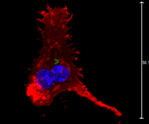
Bacille Calmette Guerin (BCG) is the most common vaccine administered to combat tuberculosis disease in the world (Figure 1). (1) The vaccine contains a weakened live strain of Mycobacterium bovis (2), which is present in cows and shares a common ancestor with the human tubercule bacillus Mycobacterium tuberculosis (3). Similar to other vaccines, BCG induces the formation of antibodies from the harmless strain of Myobacterium bovis to help prevent tuberculosis (4). Although BCG remains as the primary vaccine against tuberculosis, it has only produced variable amounts of success. Additionally, the emergence of mycobacterial drug resistance has further jeopardized the effectiveness of BCG (5). Researchers are currently searching for a more effective way to treat tuberculosis disease, but no successful vaccine is expected for twenty years (1).
Origin
The origin of the Bacile Calmette Guerin virus began when Albert Calmette and Camille Guerin started working on Mycobacterium bovis in 1908. After the stain was attenuated 230 times over thirteen years, BCG was used as a vaccine to treat tuberculosis (6). The strain went through many unknown genetic changes as it became less virulent in animals. After the original success of the vaccine, it was distributed all around the world. However, as the vaccine underwent distribution, many underwent genetic changes (2). One of these distinct changes in 1927 occurred when several genes were lost in France, and these changes continued as individual countries altered the strain to fit their own needs (7). Currently, there are seven strands that are administered throughout the world, but they differ in gene sequences and protein production (7). Note: Continue with specific genes and proteins which will lead in molecular structure and how the virus works...
Molecular Structure and Function of BCG
Problems with BCG
Conclusion and Potential New Treatments
References
1. http://www.kingcounty.gov/healthservices/health/communicable/TB/bcgvaccine.aspx 2. http://www.who.int/vaccine_research/diseases/tb/vaccine_development/bcg/en/ 3. http://whqlibdoc.who.int/hq/1996/WHO_EMC_ZOO_96.4.pdf 4. http://www.netdoctor.co.uk/infections/medicines/bcg-vaccine-ssi.html 5. http://www.who.int/immunization/wer7904BCG_Jan04_position_paper.pdf 6. http://www.plosone.org/article/info%3Adoi%2F10.1371%2Fjournal.pone.0071243 7. http://www.sciencedirect.com/science/article/pii/S096284799990206X#
Compose a title for your page. Type your exact title in the Search window, then press Go. The MicrobeWiki will invite you to create a new page with this title.
Open the class template page in "edit." Copy ALL the editable text. Then go to YOUR OWN page; edit tab. PASTE into your own page, and edit.

At right is a sample image insertion. It works for any image uploaded anywhere to MicrobeWiki. The insertion code consists of:
Double brackets: [[
Filename: PHIL_1181_lores.jpg
Thumbnail status: |thumb|
Pixel size: |300px|
Placement on page: |right|
Legend/credit: Electron micrograph of the Ebola Zaire virus. This was the first photo ever taken of the virus, on 10/13/1976. By Dr. F.A. Murphy, now at U.C. Davis, then at the CDC.
Closed double brackets: ]]
Other examples:
Bold
Italic
Subscript: H2O
Superscript: Fe3+
Edited by Scott Treiman, student of Joan Sloncewski in Biology 116 Information in Living Systems at Kenyon College
Edited by [Author Name], student of Joan Slonczewski for BIOL 116 Information in Living Systems, 2013, Kenyon College.
