Nanotubes facilitate intercellular signaling in eukaryotic and prokaryotic cells
Overview

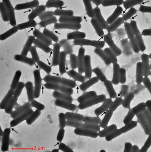
<http://www.sciencedirect.com/science?_ob=MiamiCaptionURL&_method=retrieve&_eid=1-s2.0-S009286741100016X&_image=1-s2.0-S009286741100016X-figs5.jpg&_cid=272196&_explode=defaultEXP_LIST&_idxType=defaultREF_WORK_INDEX_TYPE&_alpha=defaultALPHA&_ba=&_rdoc=1&_fmt=FULL&_issn=00928674&_pii=S009286741100016X&md5=b2d22cbeb0a19edbc6d02597fb061a4d>.
At right is a sample image insertion. It works for any image uploaded anywhere to MicrobeWiki. The insertion code consists of:
Double brackets: [[
Filename: PHIL_1181_lores.jpg
Thumbnail status: |thumb|
Pixel size: |300px|
Placement on page: |right|
Legend/credit: Electron micrograph of the Ebola Zaire virus. This was the first photo ever taken of the virus, on 10/13/1976. By Dr. F.A. Murphy, now at U.C. Davis, then at the CDC.
Closed double brackets: ]]
Other examples:
Bold
Italic
Subscript: H2O
Superscript: Fe3+
Structure
Include some current research in each topic, with at least one figure showing data.
Formation

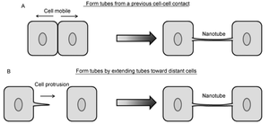
<http://link.springer.com/article/10.1007%2Fs11427-013-4548-3>.
Include some current research in each topic, with at least one figure showing data.
HIV Pathogenesis

<http://www.ncbi.nlm.nih.gov/pmc/articles/PMC2701345/figure/F5/>.
Include some current research in each topic, with at least one figure showing data.
HTLV Pathogenesis
Overall paper length should be 3,000 words, with at least 3 figures.
Prion Pathogenesis

Include some current research in each topic, with at least one figure showing data.
Prion Pathogenesis
Include some current research in each topic, with at least one figure showing data.
Genetic and cytoplasmic exchange in B. subtilis
Include some current research in each topic, with at least one figure showing data.
Metabolic mutualism between E. coli and Acinetobacter baylyi
Include some current research in each topic, with at least one figure showing data.
References
Edited by student of Joan Slonczewski for BIOL 238 Microbiology, 2009, Kenyon College.
